
You’re here because something’s not quite right. Maybe it’s that persistent ringing in your ear, a subtle imbalance, or a fullness that just won’t go away. These can be the whispers of an acoustic neuroma, a benign tumor that develops on the nerve connecting your ear to your brain. Navigating a potential diagnosis can feel overwhelming, a labyrinth of medical jargon and uncertain timelines. But fear not, because you’ve come to the right place. As your Listicle Content Architect (LCA), I’m here to break down the process of acoustic neuroma diagnosis into clear, actionable steps, empowering you with the knowledge you need. This isn’t about scaring you; it’s about equipping you. Think of this as your roadmap, guiding you from suspicion to clarity.
Your body often speaks to you long before a doctor can pinpoint an issue. For acoustic neuroma, these “whispers” can be subtle but persistent. Understanding these initial symptoms is your first crucial step in advocating for your health. Don’t dismiss them as mere annoyances; they could be vital clues.
a. The Persistent Ringing: Tinnitus as a Red Flag
You know that annoying buzz, hiss, or high-pitched whine that seems to have taken up permanent residence in your ear? That’s tinnitus, and while it has numerous causes, when it’s unilateral (affecting only one ear) and persistent, it warrants attention. It’s often one of the earliest and most common indicators of an acoustic neuroma. The tumor’s growth can put pressure on the auditory nerve, disrupting normal sound processing and leading to this phantom noise. Sometimes, it might be a low hum, other times a sharp, piercing sound. The key is its persistence and its localization to one ear. If you’ve been experiencing this for more than a few weeks, it’s time to start paying closer attention.
b. The Shifting Soundscape: Hearing Loss Demystified
Another significant sign is a gradual, often unnoticed, decline in your hearing. This usually starts with higher-pitched sounds. You might find yourself struggling to understand conversations in noisy environments, asking people to repeat themselves more often, or turning up the volume on the TV. This type of hearing loss, particularly if unilateral, is a strong indicator. The auditory nerve is responsible for transmitting sound signals, and as the neuroma grows, it can compress this nerve, impeding its ability to carry those signals effectively. It’s not like a sudden loss, but a slow erosion of your auditory world. You might not realize how much you’ve lost until you start addressing it.
c. The Disorienting Wobble: Imbalance and Dizziness Explained
Acoustic neuromas can also affect your sense of balance. This is because the vestibular nerve, responsible for maintaining equilibrium, runs very close to the auditory nerve. Pressure from the tumor can disrupt its function, leading to feelings of unsteadiness, vertigo (a sensation of spinning), or a general lack of balance. You might feel clumsy, experience difficulty walking in the dark, or find yourself struggling to maintain your posture. This symptom can be quite disconcerting and can significantly impact your daily life, making even simple tasks feel challenging. It’s a sign that something deeper might be affecting your inner ear’s complex balance system.
d. The Unsettling Fullness: Inner Ear Sensations Detailed
Beyond ringing and imbalance, some individuals report a persistent feeling of fullness or pressure within the affected ear. It’s a sensation akin to having constantly blocked ears, as you might experience during airplane ascents or descents. This can be due to the tumor’s presence and its impact on the delicate structures of the inner ear. It’s not an outward symptom you can see, but an internal sensation that can be both uncomfortable and worrying.
e. Less Common, But Not to Be Ignored: Other Potential Indicators
While the above are the most prevalent symptoms, there are other, less common indicators to be aware of. These can include:
- Facial Weakness or Numbness: In very rare cases, especially with larger tumors, the facial nerve, which runs alongside the acoustic nerve, can be affected, leading to a drooping of the face, a change in taste, or numbness on one side of the face.
- Headaches: Persistent or new-onset headaches, particularly those that are unilateral and distinct from your usual headaches, could be a cause for concern.
- Coordination Issues: Similar to imbalance, you might notice difficulties with fine motor skills or a general feeling of being uncoordinated.
2. The Doctor’s Inquiry: The Initial Medical Consultation
You’ve noticed something’s off, and you’ve bravely scheduled an appointment with your doctor. This initial consultation is where you voice your concerns and where your doctor begins to gather information. Be prepared to be an active participant in this process; your honest and detailed answers are crucial.
a. Detailing Your Symptoms: A Comprehensive History
Your doctor will start by asking you about your symptoms. This is your opportunity to be as specific as possible. Don’t downplay what you’re experiencing. Think about:
- Onset: When did the symptoms begin? Was it sudden or gradual?
- Frequency and Duration: How often do these symptoms occur, and how long do they last?
- Severity: On a scale of 1 to 10, how intense are your symptoms?
- Triggers and Alleviators: Does anything make your symptoms better or worse? For example, does noise exacerbate your tinnitus, or does lying down help with dizziness?
- Impact on Daily Life: How are these symptoms affecting your work, hobbies, and social interactions? Be honest about the challenges.
b. The Physical Examination: What to Expect
Your doctor will then perform a physical examination. This is not a definitive diagnostic test for acoustic neuroma right then and there, but it helps them rule out other conditions and assess the extent of your symptoms. This might include:
- Ear Examination: Using an otoscope to look inside your ear canal and eardrum for any visible abnormalities.
- Neurological Assessment: This is a key part. Your doctor will test your reflexes, muscle strength, coordination, balance, and gait. They might ask you to perform specific movements, like walking heel-to-toe or touching your finger to your nose.
- Hearing and Balance Screening: They might conduct a basic hearing test by whispering words or using a tuning fork. They may also assess your balance by asking you to stand with your feet together and eyes closed.
c. Ruling Out Common Culprits: Differential Diagnosis
It’s important to remember that acoustic neuroma is a relatively rare condition. Your doctor’s primary goal at this stage is to rule out more common causes of your symptoms. These could include:
- Earwax Impaction: Excessive earwax can cause muffled hearing and a feeling of fullness.
- Middle Ear Infections: These can lead to pain, hearing loss, and dizziness.
- Meniere’s Disease: This inner ear disorder can cause vertigo, tinnitus, and fluctuating hearing loss.
- Vascular Issues: In some cases, blood vessel issues can mimic certain symptoms.
d. The Referral: When Further Expertise is Needed
If your doctor suspects that your symptoms are not explained by common conditions, or if they have specific concerns based on your neurological exam, they will likely refer you to a specialist. This is usually an otolaryngologist (an ENT doctor) or a neurotologist, a physician who specializes in ear disorders and the bony structures of the skull base. This referral is a positive step, indicating that your concerns are being taken seriously and that you are moving towards more specialized diagnostic tools.
3. Unveiling the Evidence: Diagnostic Imaging Techniques

Once you’re referred to a specialist, the diagnostic process moves into the realm of advanced imaging. These powerful tools are designed to visualize the structures of your ear and brain, allowing doctors to identify or rule out the presence of an acoustic neuroma with a high degree of accuracy.
a. The Magnetic Resonance Imaging (MRI) Scan: The Gold Standard
When it comes to diagnosing acoustic neuromas, the MRI is your most powerful ally. It uses a powerful magnetic field and radio waves to create detailed, cross-sectional images of your brain and the intricate structures of your inner ear and skull base.
- Why it’s Superior: MRI excels at differentiating between different types of soft tissues, making it highly effective at visualizing the delicate nerves and any abnormal growths. It can detect even very small tumors that might be missed by other imaging methods.
- Contrast Enhancement: Often, you’ll be given an injection of a contrast dye (gadolinium) before the MRI. This dye highlights tumors and other abnormal tissues, making them appear brighter on the images and improving the clarity of the diagnosis.
- What to Expect: The MRI machine is a large, tube-like structure. You’ll lie on a table that slides into the scanner. The process can take anywhere from 30 minutes to over an hour, and it can be quite noisy. It’s important to remain as still as possible for clear images. You might be offered earplugs or headphones to help with the noise. Inform your doctor beforehand if you have any metal implants or pacemakers, as these can be contraindications for MRI.
b. The Computed Tomography (CT) Scan: A Supporting Role
While MRI is generally preferred for acoustic neuroma diagnosis due to its superior soft-tissue resolution, CT scans can sometimes play a supporting role, particularly in specific situations.
- When it Might Be Used: CT scans use X-rays to create detailed cross-sectional images. They are excellent at visualizing bone structures. A CT scan might be used to assess the bony structures of the internal auditory canal, especially if the MRI is inconclusive or if there are concerns about bony abnormalities that could be affecting the nerve.
- Limitations for Acoustic Neuroma: CT scans are not as good as MRIs at visualizing soft tissues like tumors. You might need a higher radiation dose with a CT scan compared to an MRI.
c. Interpreting the Images: The Radiologist’s Expertise
The MRI and CT scans will be meticulously reviewed by a radiologist, a physician specializing in interpreting medical images. They will look for:
- Tumor Size and Location: They will measure the size of any detected tumor and pinpoint its exact location within the internal auditory canal or extending into the cerebellopontine angle (the space at the back of the brain near the cerebellum and pons).
- Tumor Characteristics: They will assess the tumor’s shape and how it interacts with surrounding structures.
- Differential Diagnosis: They will also consider other possible explanations for any anomalies seen on the scans, ensuring a comprehensive evaluation.
4. Hearing and Balance Tests: Quantifying the Impact
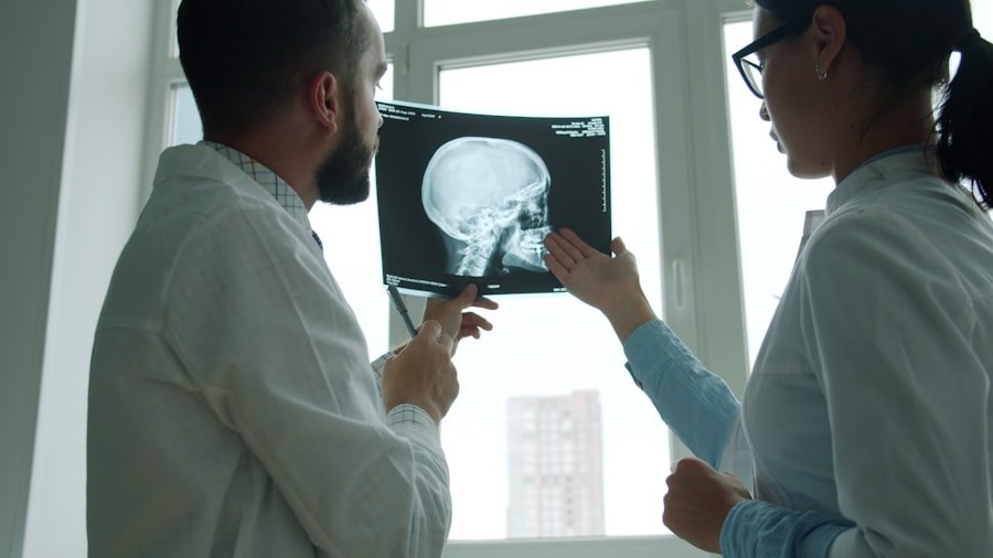
Beyond visualizing the tumor, a crucial part of the diagnostic process involves objectively assessing the functional impact of the suspected acoustic neuroma on your hearing and balance systems. These specialized tests provide quantitative data that complements the imaging results and helps determine the extent of the tumor’s influence.
a. Audiometry: Measuring Your Hearing Acuity
Audiometry is a comprehensive set of hearing tests performed by an audiologist. It’s designed to measure your ability to hear different frequencies and sound intensities.
- Pure-Tone Audiometry: This is the most common test. You’ll be in a soundproof booth and will wear headphones. You’ll be asked to raise your hand or press a button whenever you hear a tone. This helps establish your hearing threshold – the softest sound you can perceive at different pitches. For acoustic neuroma, you’ll likely see a significant loss in the higher frequencies in the affected ear.
- Speech Audiometry: This involves listening to and repeating words at different volumes. It helps assess your ability to understand speech, especially in the presence of background noise. Difficulty understanding speech, even at normal volumes, is a common finding with acoustic neuroma.
- Tympanometry: This test measures the movement of your eardrum and the pressure in your middle ear. It can help identify problems in the middle ear and rule out conditions that might mimic hearing loss.
b. Vestibular Evoked Myogenic Potentials (VEMPs): Assessing Balance Nerve Function
VEMPs are specialized tests that evaluate the function of the vestibular system, your body’s balance system. They measure nerve responses to sound stimuli.
- Cervical VEMPs (cVEMPs): These tests assess the saccule, a part of the inner ear responsible for sensing linear acceleration and tilt. You’ll hear clicking sounds through headphones, and electrodes placed on your neck will measure a muscle response.
- Ocular VEMPs (oVEMPs): These tests evaluate the utricle, another part of the inner ear that helps with sensing gravity and head movements. You’ll hear clicking sounds, and electrodes will be placed around your eyes to measure a response in your eye muscles.
- What they Reveal: Abnormal VEMP responses can indicate damage or dysfunction in the vestibular nerves, often consistent with the presence of a growing tumor that is compressing these delicate pathways.
c. Electronystagmography (ENG) or Videonystagmography (VNG): Mapping Eye Movements for Balance Clues
| Diagnostic Test | Accuracy | Advantages | Disadvantages |
|---|---|---|---|
| MRI | High | Non-invasive, detailed imaging | Expensive |
| CT Scan | Lower than MRI | Quick results | Less detailed |
| Hearing Test | Indirect | Assess hearing loss | Does not directly detect tumor |
ENG and VNG are tests that record the movements of your eyes, which are intimately linked to your sense of balance.
- How it Works: You’ll wear special goggles with cameras that track your eye movements. Your doctor might then introduce different stimuli, such as:
- Tracking moving lights: To assess your ability to follow visual targets.
- Following your own finger: To check smooth pursuit eye movements.
- Rapid eye movements: To test saccadic function.
- Introducing cold and warm air or water into your ear canals: This is a critical part, known as caloric testing. It stimulates the vestibular system and causes involuntary eye movements called nystagmus. If one ear’s vestibular system is significantly impaired by a tumor, the response to this stimulation will be reduced or absent.
- Interpreting the Data: The recorded eye movements provide valuable information about the health of your vestibular system and can help pinpoint any imbalance or dizziness originating from the inner ear.
d. Brainstem Auditory Evoked Response (BAER) or Auditory Brainstem Response (ABR): Measuring Nerve Signal Transit
BAER, also known as ABR, is a non-invasive test that measures the electrical activity in the auditory nerve and brainstem pathways in response to sound.
- The Procedure: Small electrodes are placed on your scalp and earlobes. You’ll wear headphones that deliver clicking sounds. The electrodes detect the brainwave responses as the sound travels from the ear through the auditory nerve to the brainstem.
- What it Shows: For an acoustic neuroma, the time it takes for the signal to travel through the affected auditory nerve will be significantly prolonged, or the response may be absent altogether. This test is particularly sensitive to abnormalities in the nerve pathway and can help confirm the presence of a tumor impacting nerve function.
5. Diagnosis Confirmation and the Road Ahead: Understanding Your Results
You’ve undergone imaging, undergone hearing and balance tests, and now it’s time for the moment of truth: receiving your diagnosis. This step is about understanding what the results mean and what the next steps will be in managing your health.
a. Bringing All the Pieces Together: The Specialist’s Evaluation
Your ENT specialist or neurotologist will synthesize all the information gathered from your medical history, physical examination, imaging scans, and functional tests. They will use this comprehensive picture to:
- Confirm or Rule Out Acoustic Neuroma: Based on the evidence, they will definitively state whether an acoustic neuroma is present.
- Determine Tumor Size and Characteristics: They will provide you with precise details about the tumor’s size, its exact location, and any specific features that might influence treatment decisions.
- Assess the Impact on Your Hearing and Balance: They will explain how the tumor is affecting your auditory and vestibular functions based on the test results.
b. Discussing Treatment Options: A Personalized Approach
If an acoustic neuroma is confirmed, your doctor will discuss the available treatment options. It’s important to understand that acoustic neuromas often grow very slowly, and not all require immediate surgical intervention. Your options will be tailored to your specific situation, considering factors such as:
- Tumor Size: Smaller tumors may be monitored, while larger ones may require treatment.
- Rate of Growth: If imaging shows the tumor is growing rapidly, treatment might be recommended sooner.
- Your Age and Overall Health: Your general health status will influence the suitability of different treatment approaches.
- Severity of Your Symptoms: The impact of the tumor on your hearing, balance, and other functions will be a significant factor.
- Your Personal Preferences: Your doctor will listen to your concerns and preferences to collaboratively decide on the best course of action.
c. Potential Treatment Pathways: A Spectrum of Possibilities
The primary treatment approaches for acoustic neuroma include:
- Observation (Watchful Waiting): For small, slow-growing tumors with minimal symptoms, regular MRI scans and audiometric evaluations might be the best course of action. This approach allows you to avoid the risks and side effects of treatment while closely monitoring the tumor’s progression.
- Surgery: This is often recommended for larger tumors, tumors that are growing rapidly, or those causing significant symptoms. The goal is to remove the tumor entirely. Common surgical approaches include:
- Middle Fossa Craniotomy: For tumors located near the inner ear.
- Retrosigmoid Approach: For tumors larger than those treated with the middle fossa approach.
- Translabyrithine Approach: Primarily used when hearing preservation is not a goal.
Your surgeon will discuss the specific risks and benefits of each surgical technique related to your tumor.
- Radiation Therapy (Radiosurgery): This involves precisely focused beams of radiation to target and shrink or stop the growth of the tumor without surgically removing it. Techniques like Stereotactic Radiosurgery (SRS), such as Gamma Knife or CyberKnife, are highly precise and can be an effective option, particularly for tumors where surgery might be more complicated or carry higher risks.
d. Embracing the Journey: Support and Information
Receiving a diagnosis can bring a mix of emotions. It’s natural to feel anxious, uncertain, or even relieved to finally have an answer. Remember, you are not alone in this journey.
- Seek Support: Connect with family, friends, and support groups for individuals with acoustic neuroma. Sharing experiences and concerns can be incredibly validating and empowering.
- Ask Questions: Don’t hesitate to ask your doctor every question you have, no matter how small it may seem. Understanding your diagnosis and treatment plan thoroughly is crucial for peace of mind.
- Educate Yourself: Continue to learn about acoustic neuroma and your treatment options. Reliable sources of information can help you feel more in control.
This guide has illuminated the path from recognizing potential symptoms to understanding a confirmed diagnosis. You’ve been equipped with the knowledge to navigate this complex process, empowering you to be an active and informed participant in your healthcare. Remember, your proactive engagement is your greatest asset.
FAQs
What is an acoustic neuroma?
An acoustic neuroma is a non-cancerous tumor that develops on the main nerve leading from the inner ear to the brain. It is also known as a vestibular schwannoma.
What are the symptoms of acoustic neuroma?
Symptoms of acoustic neuroma may include hearing loss, ringing in the ear, dizziness, balance problems, facial numbness or weakness, and headaches.
How is acoustic neuroma diagnosed?
Acoustic neuroma can be diagnosed through a combination of medical history, physical examination, hearing tests, and imaging studies such as MRI or CT scans.
What are the treatment options for acoustic neuroma?
Treatment options for acoustic neuroma may include observation, radiation therapy, or surgical removal of the tumor, depending on the size and location of the tumor and the patient’s overall health.
What are the potential complications of acoustic neuroma?
Complications of acoustic neuroma may include permanent hearing loss, facial weakness or paralysis, balance problems, and damage to nearby nerves and blood vessels during surgical removal.