
You’re a curious individual, and you’ve found yourself wading into the complex waters of hearing loss, specifically when it intersects with a rather unusual condition: acoustic neuroma. As the Listicle Content Architect, your mission is to distill this intricate topic into easily digestible, actionable insights. No jargon overload, just pure, focused understanding. Let’s dive in.
You’ve heard the term, maybe even seen it in a medical context, but what is an acoustic neuroma? Let’s break it down, layer by layer.
1.1 The “Neuroma” Part: A Tumor, But What Kind?
The “neuroma” in acoustic neuroma tells you it’s a type of tumor. But it’s not just any tumor. It’s specifically a benign tumor, meaning it’s not cancerous and doesn’t spread to other parts of the body. Its origin is key to understanding its name.
- Schwann Cells: Your nerves are wrapped in a protective fatty layer called myelin, much like insulation on electrical wires. This myelin sheath is produced by specialized cells called Schwann cells. Acoustic neuromas arise from these Schwann cells.
- Vestibular Schwannoma: You might also hear acoustic neuroma referred to as a vestibular schwannoma. This term is more precise because it pinpoints the specific nerve involved: the vestibular nerve.
1.2 The “Acoustic” Part: The Nerve in Question
The “acoustic” refers to the vestibulocochlear nerve, which is critically important for two of your senses:
- Hearing (Cochlear Nerve): This branch of the nerve transmits sound signals from your inner ear to your brain, allowing you to perceive what you hear.
- Balance (Vestibular Nerve): This branch is responsible for your sense of equilibrium and spatial orientation. It tells your brain where your head is in space and how it’s moving.
So, an acoustic neuroma is a benign tumor that develops on the vestibulocochlear nerve, typically on the myelin sheath of the vestibular division.
1.3 How and Where Does It Grow?
These tumors usually grow very slowly, often over years or even decades. They emerge from the internal auditory canal, a narrow bony passage in your skull that houses the vestibulocochlear nerve, the facial nerve (which controls facial muscles), and blood vessels.
- Location, Location, Location: Because the internal auditory canal is a confined space, even a small tumor can begin to press on the delicate nerves housed within it. As it grows, it can extend further into the cerebellopontine angle, a space at the base of the brain where cranial nerves emerge.
- Gradual Onset: The slow growth means that symptoms often develop gradually. This can sometimes make diagnosis challenging, as early symptoms might be dismissed as normal age-related changes or minor inconveniences.
1.4 Is It Common? And Who Gets It?
Acoustic neuromas are considered rare tumors. They account for about 8% of all tumors found within the skull.
- Incidence: The incidence is estimated to be around 1 to 3 new cases per 100,000 people each year. This means you’re unlikely to encounter someone with acoustic neuroma in your daily life, but for those affected, it’s a significant health concern.
- Age and Gender: They most commonly affect adults between the ages of 30 and 60. While they can occur at any age, this age range is when they are most frequently diagnosed. There isn’t a significant gender predisposition; they affect men and women roughly equally.
- Genetic Factors (Neurofibromatosis Type 2): In rare instances (about 5% of cases), acoustic neuromas occur as part of a genetic disorder called Neurofibromatosis Type 2 (NF2). NF2 causes tumors to develop on nerve tissues throughout the body, and acoustic neuromas are a hallmark of this condition. If you have a family history of NF2 or develop bilateral acoustic neuromas (one on each side), genetic testing might be recommended.
2. The Domino Effect: How Acoustic Neuromas Cause Hearing Loss and Other Symptoms
You’re experiencing a change in your hearing, and you’re wondering how a tiny tumor could be the culprit. The answer lies in how the acoustic neuroma impacts the delicate structures it grows upon. Think of it like a slow, steady pressure that gradually disrupts essential functions.
2.1 The Gradual Squeeze: Pressure on the Cochlear Nerve
The cochlear nerve, your hearing pathway, is particularly vulnerable. As the acoustic neuroma enlarges, it exerts increasing pressure on this nerve. This pressure doesn’t instantly silence your hearing; instead, it interferes with the transmission of nerve signals.
- Distortion of Sound: The first sign isn’t usually complete deafness. It’s more often a subtle distortion of sound. You might notice that speech sounds muffled or unclear, especially in noisy environments.
- Monaural Hearing Loss: A key characteristic of acoustic neuroma-related hearing loss is that it typically affects one ear only (unilateral). This is because the tumor is usually unilateral. If you experience hearing loss in both ears that’s related to an acoustic neuroma, it strongly suggests NF2.
- Progressive Nature: This hearing loss is almost always progressive. It doesn’t usually come on suddenly. You might notice it worsening over months or years, making it easy to adapt to initially, but ultimately leading to significant impairment.
- Frequency Specificity: Often, the hearing loss starts with higher frequencies. This means you might struggle to hear the ‘s’, ‘f’, and ‘th’ sounds, which are crucial for distinguishing words. This can lead to mishearing or simply not understanding what’s being said.
2.2 The Unwanted Soundtrack: Tinnitus as an Early Warning
You’ve noticed a ringing, buzzing, or humming sound in your ear – a constant companion that shouldn’t be there. This is tinnitus, and for many people with acoustic neuroma, it’s one of the earliest and most noticeable symptoms.
- Where Does It Come From? The pressure and irritation on the cochlear nerve by the growing tumor are believed to be the cause of the tinnitus. The nerve is essentially sending false signals to the brain, which you perceive as sound.
- Constant and Unilateral: Like the hearing loss, tinnitus associated with acoustic neuroma is typically constant and in the affected ear. The pitch and loudness can vary, but it’s a persistent presence.
- Impact on Daily Life: Tinnitus can be incredibly disruptive. It can interfere with sleep, concentration, and overall mood, leading to anxiety and frustration.
2.3 Losing Your Balance: When the Vestibular Nerve is Affected
The vestibular nerve is your internal gyroscope. When a tumor presses on it, your sense of balance is directly compromised. This can manifest in a variety of unsettling ways.
- Dizziness and Vertigo: This is a hallmark symptom. You might experience dizziness, a vague feeling of unsteadiness, or vertigo, a more intense sensation that you or your surroundings are spinning.
- Imbalance and Falls: The impaired balance can lead to a feeling of unsteadiness when walking, making you more prone to stumbling or falling. This can be particularly concerning for older adults.
- Nausea and Vomiting: In severe cases of vertigo, nausea and even vomiting can occur, similar to motion sickness.
- The “Tip-Toe” Gait: Some individuals develop a slight alteration in their walking style, often unconsciously, as they try to maintain better balance. They might feel more stable when walking with their feet slightly closer together or even on their toes.
2.4 Other Nerves in the Crossfire: When the Tumor Grows Larger
Acoustic neuromas typically start small and grow outwards. As they increase in size and extend into the cerebellopontine angle, they can begin to compress other important cranial nerves.
- Facial Nerve (Cranial Nerve VII): This nerve controls the muscles of your face. If compressed, you might experience:
- Facial Weakness or Numbness: This can range from a slight droop on one side of your face to difficulty closing your eye or smiling.
- Taste Disturbances: In some cases, the tumor can affect the taste sensation on the front of the tongue.
- Trigeminal Nerve (Cranial Nerve V): This nerve is responsible for sensation in your face and functions related to chewing. Compression can lead to:
- Facial Numbness or Tingling: You might notice a loss of sensation or a pins-and-needles feeling on one side of your face.
- Jaw Pain or Difficulty Chewing: In rarer cases, pain or discomfort in the jaw may occur.
- Brainstem Compression (Very Large Tumors): In extremely rare and advanced cases, a very large tumor can compress the brainstem, the vital structure connecting the brain to the spinal cord. This can lead to more severe neurological symptoms, including headaches, swallowing difficulties, and even changes in consciousness. Fortunately, with modern diagnostics, tumors are usually detected long before this stage.
3. Unmasking the Culprit: Diagnostic Pathways for Acoustic Neuroma
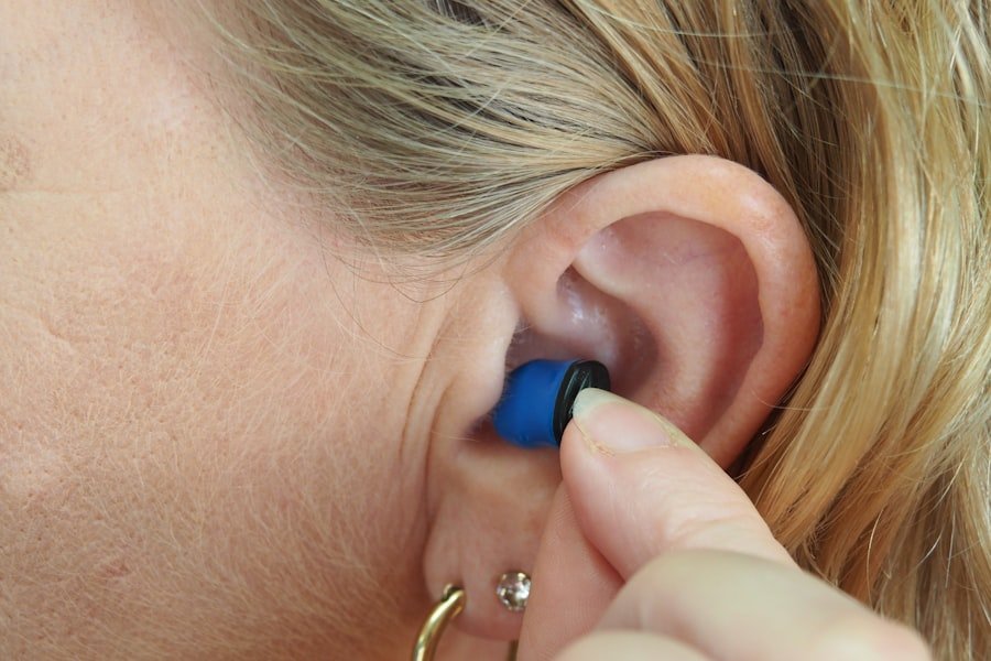
You’ve been experiencing some concerning symptoms, and the thought of an acoustic neuroma has crossed your mind. Now, you’re wondering how doctors pinpoint this condition. The diagnostic journey involves a combination of careful listening to your story, physical examinations, and sophisticated imaging techniques.
3.1 The Initial Clues: Patient History and Physical Examination
Your doctor’s first and most crucial step is to gather information from you and to conduct a thorough physical examination.
- The Power of Your Story: Be prepared to discuss your symptoms in detail. When did they start? How have they progressed? Are they constant or intermittent? Which ear is affected? Your detailed account is invaluable.
- Hearing Tests (Audiometry): This is a fundamental part of the investigation.
- Pure-Tone Audiometry: This test measures your ability to hear sounds at different frequencies and intensities. In acoustic neuroma, you’ll typically see a pattern of hearing loss, often greater in the higher frequencies and affecting only one ear.
- Speech Audiometry: This assesses your ability to understand spoken words. You might find it more difficult to comprehend speech, especially in the presence of background noise, even if you can hear certain tones.
- Tests of Balance and Neurological Function: Your doctor will also assess your balance, coordination, and the function of other cranial nerves. This might involve checking your eye movements, facial muscle strength, and reflexes.
3.2 Peering Inside: Imaging Techniques
Once the initial suspicions are raised, imaging becomes essential for visualizing the tumor and confirming its presence.
- Magnetic Resonance Imaging (MRI): The Gold Standard: This is the most accurate and commonly used imaging technique for diagnosing acoustic neuroma.
- How it Works: MRI uses powerful magnets and radio waves to create detailed cross-sectional images of your brain and the internal auditory canals.
- Contrast Enhancement: Often, a contrast dye (gadolinium) is injected into your bloodstream before the MRI. This dye highlights the tumor, making it much easier to see against the surrounding tissues. Acoustic neuromas typically enhance brightly with contrast.
- Why it’s Superior: MRI provides exceptional detail of soft tissues, allowing doctors to precisely measure the size and location of the tumor and assess its impact on nearby structures.
- Computed Tomography (CT) Scan: A Complementary Tool: While MRI is preferred, a CT scan can sometimes be used, especially if MRI is not an option or to provide additional information about the bone structure of the internal auditory canal.
- How it Works: CT scans use X-rays to create detailed images.
- Bone Evaluation: CT is particularly useful for visualizing bone abnormalities that might be associated with or contributing to nerve issues. However, it’s less effective at visualizing the soft tissue of the tumor itself compared to MRI.
3.3 Confirming a Diagnosis: Beyond Imaging
While imaging is key, sometimes further tests are used to gather more information or rule out other conditions.
- Auditory Brainstem Response (ABR) Testing: This electrophysiological test measures the electrical activity in your auditory nerve and brainstem in response to sound.
- How it Works: Small sensors are placed on your scalp and earlobes, and you listen to clicking sounds. The ABR measures how quickly and effectively the auditory signals travel from the ear to the brainstem.
- Indications: In acoustic neuroma, the ABR will show delays in signal transmission, particularly on the affected side, indicating a problem with the nerve pathway. It can even detect very small tumors that might be missed by MRI in rare instances.
- Genetic Testing (for NF2): If there is a suspicion of Neurofibromatosis Type 2, perhaps due to bilateral acoustic neuromas or a family history, genetic testing can definitively diagnose the condition. This involves analyzing a sample of your blood or saliva for specific genetic mutations.
4. Your Options for Action: Treatment Strategies for Acoustic Neuroma
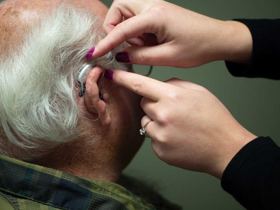
You’ve been diagnosed with an acoustic neuroma, and now the question on your mind is: what happens next? The good news is that there are several effective treatment approaches, and the best path forward depends on a variety of factors.
4.1 The Watchful Waiting Game: Observation and Monitoring
For very small tumors that are not causing significant symptoms, a strategy of watchful waiting might be recommended. This doesn’t mean you’re being ignored; it means your doctor believes the risks of immediate intervention outweigh the benefits.
- When is Observation Appropriate? This approach is typically considered for tumors that are:
- Very Small: Generally less than 1-2 cm in diameter.
- Asymptomatic or Minimally Symptomatic: Not causing significant hearing loss, balance problems, or other troubling symptoms.
- Slow-Growing: Based on imaging history, it appears to be growing very slowly or not at all.
- Regular Monitoring: If you’re on a watch-and-wait plan, you’ll need to undergo regular follow-up appointments and MRI scans to monitor the tumor’s size and growth. This allows for early detection of any progression.
- The Goal: The goal of observation is to avoid or delay the potential side effects of surgery or radiation while ensuring that any changes in the tumor are caught in time for treatment.
4.2 Precision Strikes: Radiation Therapy Options
Radiation therapy uses targeted beams of high-energy radiation to damage and destroy tumor cells or to stop them from growing. For acoustic neuroma, this is often a non-invasive or minimally invasive option.
- Stereotactic Radiosurgery (SRS): The Most Common Method: This is a highly precise form of radiation therapy.
- How it Works: Multiple beams of radiation are delivered from different angles, converging on the tumor with extreme accuracy, minimizing damage to surrounding healthy tissues.
- Key Techniques: Popular methods include Gamma Knife, CyberKnife, and linear accelerator-based stereotactic radiosurgery.
- Advantages: SRS is typically performed in one or a few outpatient sessions. It’s excellent for controlling tumor growth and often preserves hearing and facial nerve function better than surgery for certain tumor sizes and locations.
- Fractionated Radiotherapy: In some cases, particularly for larger tumors or when there are concerns about damaging critical structures, radiation might be delivered in smaller doses over several weeks. This is known as fractionated radiotherapy.
- Goals of Radiation: The primary goal of radiation is to stop the tumor from growing further. It may not necessarily shrink the tumor significantly, but halting its progression is often sufficient to alleviate symptoms and prevent further damage.
4.3 Surgical Intervention: Removing the Tumor
For larger tumors, tumors that are growing rapidly, or those causing significant symptoms, surgery to remove the acoustic neuroma may be the recommended course of action.
- Types of Surgical Approaches: The surgeon will choose an approach based on the tumor’s size, location, and proximity to important nerves.
- Retrosigmoid Approach: This is a common approach where an incision is made behind the ear, and the surgeon accesses the tumor through the space behind the brainstem. It offers good visualization of the tumor and nerves.
- Translabyrinthine Approach: This approach involves removing part of the bone behind the ear and traversing through the inner ear structures. It provides excellent access to the tumor but typically results in complete hearing loss in the operated ear and also affects balance.
- Middle Fossa Approach: This approach involves an incision above the ear and is often used for smaller tumors located higher up in the internal auditory canal. It may offer a better chance of preserving hearing.
- Goals of Surgery: The aim of surgery is to remove as much of the tumor as possible, ideally completely.
- Potential Risks and Side Effects: Surgery carries inherent risks, including:
- Hearing Loss: Even with the most skilled surgeons, some degree of hearing loss in the affected ear is common, especially in larger tumors.
- Facial Nerve Weakness or Paralysis: The facial nerve is very close to the tumor, and there’s a risk of injury, leading to temporary or permanent weakness or paralysis of facial muscles.
- Balance Problems: Surgery can affect the balance system, leading to temporary or persistent dizziness.
- Cerebrospinal Fluid (CSF) Leak: A rare complication where the fluid surrounding the brain and spinal cord leaks out.
- Tinnitus: While the tumor might be removed, tinnitus can sometimes persist or even worsen post-surgery.
- Microsurgery: Surgeons use microscopes and specialized instruments to perform these delicate procedures, enhancing precision and minimizing damage.
4.4 Post-Treatment Considerations: Life After Acoustic Neuroma Treatment
| Category | Data/Metrics |
|---|---|
| Prevalence of Hearing Loss | Approximately 15% of adults aged 18 and over report some trouble hearing |
| Types of Hearing Loss | Conductive, Sensorineural, and Mixed |
| Acoustic Neuroma Incidence | 1 in 100,000 people per year |
| Acoustic Neuroma Symptoms | Hearing loss, tinnitus, balance problems, and facial numbness |
| Treatment Options | Monitoring, surgery, and radiation therapy |
Regardless of the treatment chosen, follow-up care is crucial, and you’ll likely have ongoing management needs.
- Regular Follow-Up: You’ll need to attend regular medical appointments and have periodic MRI scans to ensure the tumor hasn’t returned or regrown.
- Hearing Rehabilitation: If you’ve experienced hearing loss, options like hearing aids, cochlear implants (in select cases), or assistive listening devices can significantly improve your ability to communicate.
- Balance Rehabilitation: Physical therapy can be very beneficial for managing dizziness and improving balance and coordination.
- Tinnitus Management: Various techniques, including sound therapy, cognitive behavioral therapy (CBT), and lifestyle adjustments, can help manage the impact of tinnitus.
- Emotional and Psychological Support: Dealing with a diagnosis like acoustic neuroma and its after-effects can be emotionally taxing. Support groups and counseling can provide valuable emotional resilience.
5. Living Well: Navigating Life with Hearing Loss and Acoustic Neuroma Concerns
You’ve faced a diagnosis, and you’re navigating a new reality. Living with hearing loss, especially when it’s linked to a condition like acoustic neuroma, requires a proactive and adaptive approach. The good news is that with the right strategies and support, you can continue to lead a full and engaging life.
5.1 Empowering Your Communication: Strategies for Hearing Loss
Hearing loss doesn’t have to be a barrier to connection. You can reclaim your ability to communicate effectively with a few thoughtful adjustments.
- Strategic Seating: In conversations or group settings, try to position yourself so you can see everyone’s faces. This allows you to utilize visual cues like lip-reading and facial expressions.
- Reduce Background Noise: Whenever possible, choose quieter environments for conversations. If you can’t avoid noise, turn off distracting appliances or TV.
- Ask for Repetition and Clarification: Don’t be afraid to say “Could you please repeat that?” or “I didn’t quite catch that.” Most people are happy to oblige. You can also ask them to rephrase it.
- Face Your Speaker: Turn your head towards the person who is speaking. This ensures you are receiving the sound as directly as possible and allows you to see their mouth.
- Utilize Hearing Technology:
- Hearing Aids: Modern hearing aids are sophisticated devices that can amplify sounds and, in some cases, help with directional hearing. It’s crucial to get properly fitted by an audiologist.
- Assistive Listening Devices (ALDs): These devices can be used in conjunction with or independently of hearing aids. Examples include personal amplifiers, FM systems (often used in classrooms or larger groups), and certain TV listening devices.
- Sign Language and Captioning: Consider learning basic sign language if you’re facing significant hearing loss. Also, utilize closed captions on television and online videos. Many video conferencing platforms now offer live captioning.
5.2 Managing Dizziness and Balance Issues
The impact of acoustic neuroma on your balance can be disorienting. However, there are ways to mitigate these challenges and improve your stability.
- Vestibular Rehabilitation Therapy (VRT): This is a specialized form of physical therapy designed to help your brain compensate for problems in your inner ear. VRT exercises are tailored to your specific needs and can improve gaze stability, balance, and reduce dizziness.
- Home Safety Modifications: To prevent falls, make your home as safe as possible:
- Clear Pathways: Ensure walkways are free of clutter.
- Adequate Lighting: Ensure all areas are well-lit, especially stairs and hallways.
- Non-Slip Surfaces: Use non-slip mats in bathrooms and kitchens.
- Handrails: Install handrails on stairs and in bathrooms.
- Pacing Yourself: Avoid sudden movements or activities that exacerbate your dizziness. Break down tasks into smaller, more manageable steps.
- Hydration and Diet: Staying well-hydrated is important. Some individuals find that avoiding caffeine, alcohol, and excessive salt can help manage dizziness.
- Medical Management: In some cases, your doctor may prescribe medication to help alleviate acute dizziness or nausea.
5.3 Coping with Tinnitus: Strategies for Quieting the Noise
Tinnitus can be a constant, unwelcome companion. While it might not be entirely eliminated, you can learn to manage its impact and reduce its intrusiveness.
- Sound Therapy: Introducing gentle background sounds can help mask the tinnitus. This can include white noise machines, nature sounds, or soft music. Many people find relief by having a fan or radio on at a low volume in their bedroom at night.
- Cognitive Behavioral Therapy (CBT): CBT is a form of therapy that helps you change your thoughts and behaviors related to tinnitus. It can reduce the distress and anxiety associated with the sound, making it less bothersome.
- Stress Management: Stress often exacerbates tinnitus. Practicing relaxation techniques like meditation, deep breathing exercises, or yoga can be beneficial.
- Limit Exposure to Loud Noises: Protecting your hearing from loud noises is crucial. Wear earplugs in noisy environments like concerts or when using machinery.
- Avoid Triggers: Some individuals find that certain substances like caffeine, nicotine, and alcohol can worsen their tinnitus. Pay attention to what seems to affect you personally.
- Focus on Well-being: Engage in activities you enjoy and maintain a healthy lifestyle. Tinnitus can be less noticeable when you are feeling generally well and engaged.
5.4 Building Your Support Network: Emotional and Practical Assistance
You don’t have to go through this alone. Connecting with others and seeking professional support can make a significant difference.
- Support Groups: Connecting with others who have acoustic neuroma or similar conditions can provide a sense of community, shared experiences, and valuable coping strategies. Look for local or online support groups.
- Family and Friends: Openly communicate your needs and challenges to your loved ones. Their understanding and practical help can be invaluable.
- Mental Health Professionals: If you are experiencing anxiety, depression, or difficulty coping with the emotional impact of your diagnosis or treatment, consider seeking support from a therapist or counselor.
- Patient Advocacy Organizations: Organizations dedicated to acoustic neuroma or brain tumors can offer resources, information, and connect you with experts and support networks.
Your journey with acoustic neuroma and hearing loss is unique, but by understanding the condition, exploring your treatment options, and adopting effective management strategies, you can continue to live a vibrant and fulfilling life. Remember, knowledge is power, and you are in the driver’s seat of your well-being.
FAQs
What is acoustic neuroma?
Acoustic neuroma, also known as vestibular schwannoma, is a non-cancerous tumor that develops on the main nerve leading from the inner ear to the brain. It can cause hearing loss, ringing in the ear, and unsteadiness.
What are the symptoms of acoustic neuroma?
Symptoms of acoustic neuroma may include hearing loss, ringing in the ear, unsteadiness, dizziness, and facial numbness or weakness. These symptoms can vary depending on the size and growth rate of the tumor.
How does acoustic neuroma cause hearing loss?
Acoustic neuroma can cause hearing loss by putting pressure on the auditory nerve, which is responsible for transmitting sound signals from the inner ear to the brain. This pressure can lead to a gradual loss of hearing in the affected ear.
How is acoustic neuroma diagnosed?
Acoustic neuroma is typically diagnosed through a combination of hearing tests, imaging studies such as MRI or CT scans, and a thorough medical history and physical examination. These tests can help determine the size and location of the tumor.
What are the treatment options for acoustic neuroma?
Treatment options for acoustic neuroma may include observation, radiation therapy, or surgical removal of the tumor. The choice of treatment depends on factors such as the size and growth rate of the tumor, as well as the patient’s overall health and preferences.