You’ve been diagnosed with an acoustic neuroma, a non-cancerous tumor on the nerve that connects your ear to your brain. It’s a lot to take in, we know. The good news is that in many cases, especially for smaller tumors or when symptoms are mild, surgery isn’t the immediate go-to. Instead, a common and often highly effective approach is “watchful waiting,” also known as active surveillance or monitoring. You’re probably wondering what this entails, how it works, and what you can expect. As your Listicle Content Architect, I’m here to break it down for you, ensuring you understand every facet of monitoring your acoustic neuroma without the need for surgery, and empowering you with the knowledge to navigate this journey confidently.
This isn’t about ignoring a problem; it’s about intelligently tracking it, making informed decisions, and prioritizing your well-being. We’ll delve into the reasons behind choosing monitoring, the specific tests and evaluations involved, the crucial role of your medical team, and how you can actively participate in your care. So, settle in, and let’s explore this less invasive but incredibly important path forward.
You might be asking, “Why monitor? Shouldn’t we just deal with it?” It’s a valid question, and the answer lies in understanding the nature of acoustic neuromas and the potential downsides of immediate surgical intervention. Not all acoustic neuromas behave the same way, and for many, growth is slow, or they may not grow at all. This is precisely where the strategy of monitoring shines.
The Slow-Growing Nature of Most Acoustic Neuromas
Acoustic Neuromas Are Typically Benign
This is perhaps the most critical factor. Acoustic neuromas are a type of tumor that arises from the Schwann cells, which are responsible for the protective myelin sheath around nerves. Importantly, they are almost always benign, meaning they do not spread to other parts of the body. While they can cause symptoms by pressing on surrounding nerves and structures, their inherent nature is not aggressive or life-threatening in the same way as malignant cancers. This inherent benign nature allows for a less urgent, more observational approach.
Size and Location as Key Determinants
Your tumor’s size and precise location play a significant role in the decision to monitor. Smaller tumors, especially those less than 2-3 centimeters in diameter, are often prime candidates for monitoring. If the tumor isn’t causing significant symptoms or pressing on vital structures like the brainstem, the risks associated with immediate surgery might outweigh the benefits. Doctors will meticulously assess the tumor’s dimensions and its proximity to critical nerves and blood vessels, like the facial nerve (responsible for facial movement) and the vestibulocochlear nerve (responsible for hearing and balance). The closer it is to these delicate structures, the higher the stakes involved with any intervention.
Individual Symptom Presentation and Severity
Your experience is paramount. If your acoustic neuroma is discovered incidentally during an imaging scan for another reason, and you are experiencing minimal or no symptoms, monitoring becomes an even more appealing option. Conversely, if you are experiencing significant hearing loss, tinnitus, dizziness, or facial numbness, the decision might lean towards other treatment modalities sooner. However, even with symptoms, if they are manageable and the tumor’s growth potential is deemed low, monitoring can still be the chosen path, allowing for a period of observation before committing to more invasive treatments.
Minimizing Risks Associated with Surgery and Radiation
Surgery, while often highly effective, carries inherent risks. These can include hearing loss (often unavoidable even with the best surgical techniques), facial nerve damage, balance issues, headaches, and in rare cases, more serious complications. Radiation therapy, another treatment option, also has its own set of potential long-term side effects. Monitoring, by definition, aims to avoid these risks altogether, at least initially. If the tumor remains stable, you bypass the potential complications entirely. This focus on preserving quality of life and minimizing intervention is a cornerstone of the monitoring approach.
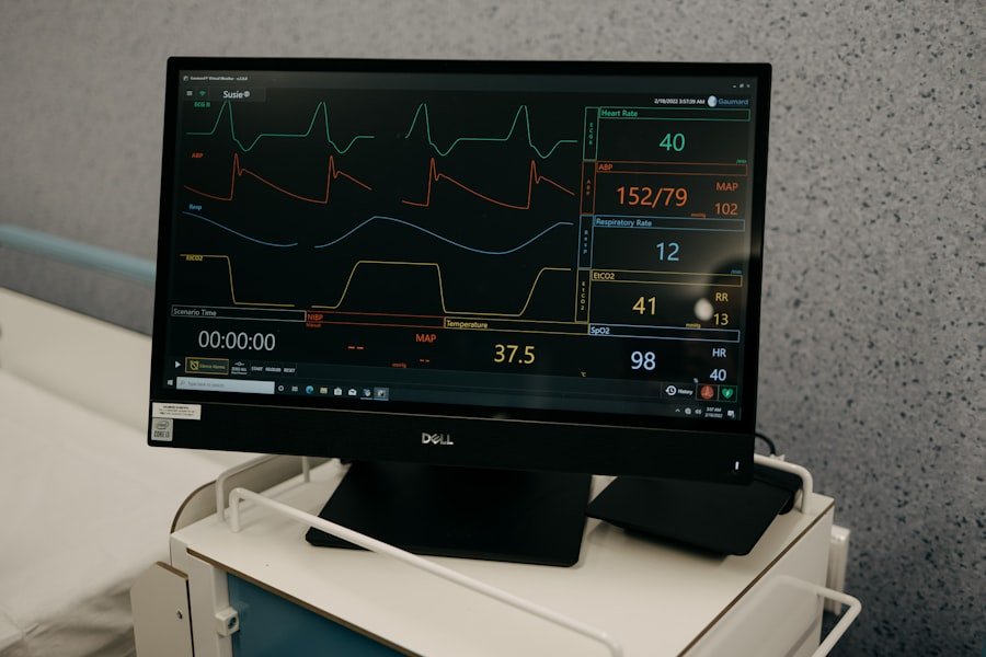
The Comprehensive Monitoring Strategy: What to Expect
So, monitoring isn’t just a passive waiting game; it’s an active and systematic process involving regular check-ups and advanced imaging techniques. You’ll be working closely with your medical team, and your commitment to attending these appointments is crucial for the success of this strategy.
Regular Audiological Examinations
These are your ears’ report cards. Audiological tests are fundamental to monitoring an acoustic neuroma because hearing loss is a common symptom. You can expect a battery of tests to be performed periodically.
Pure-Tone Audiometry
This familiar test measures your ability to hear different frequencies at varying intensities. It helps quantify the degree and type of hearing loss you might be experiencing, and importantly, can detect subtle changes over time that might indicate tumor growth or pressure on the auditory nerve. You’ll be asked to raise your hand or press a button when you hear a sound.
Speech Discrimination Testing
This goes beyond simply hearing a tone; it assesses how well you understand spoken words. You’ll be asked to repeat words or phrases at different volumes. A decline in speech discrimination can be a sensitive indicator of nerve damage or compression, even if your overall hearing levels haven’t changed drastically.
Tympanometry
This test measures the function of your middle ear and the mobility of your eardrum. It helps identify any issues with the eardrum or the small bones in the middle ear, which can sometimes be indirectly affected or contribute to your symptoms.
Otoacoustic Emissions (OAEs)
OAEs are subtle sounds produced by the inner ear’s sensory cells (hair cells). They are a quick and non-invasive way to assess the health of the cochlea. A reduction or absence of OAEs can indicate damage to these delicate hair cells, which are part of the auditory pathway.
Advanced Imaging Techniques: Your Tumor’s Blueprint
Imaging is the eyes of the monitoring process, allowing your doctors to visualize your tumor and track its size and any potential changes.
Magnetic Resonance Imaging (MRI)
MRI is the gold standard for monitoring acoustic neuromas. It provides detailed, three-dimensional images of the brain and its structures, including the internal auditory canal where the tumor resides.
Gadolinium-Enhanced MRI
Most acoustic neuroma monitoring MRIs will involve the use of a contrast agent called gadolinium. This substance is injected into your bloodstream and highlights the tumor, making it appear brighter on the scan. This enhanced visualization is crucial for accurately measuring the tumor’s dimensions and detecting even small changes in its size.
Frequency and Timing of MRIs
The frequency of your MRIs will be determined by your doctor based on factors like the tumor’s initial size, its growth rate (if known from previous scans), your symptoms, and your age. Initially, you might have MRIs every six months to a year. If the tumor remains stable over several years, the interval between scans might be extended to two or even three years.
Other Imaging Modalities (Less Common for Routine Monitoring)
While MRI is dominant, other imaging techniques might be used in specific circumstances, though they are not typically part of routine monitoring.
Computed Tomography (CT) Scans
CT scans use X-rays to create cross-sectional images. While less detailed than MRI for soft tissues like nerves, they can be useful for visualizing bone structure and are sometimes used if an MRI is contraindicated (e.g., due to metal implants).
Vestibular and Balance Testing
Since the vestibulocochlear nerve also governs balance, changes in this area are closely monitored.
Videonystagmography (VNG)
VNG is a series of tests that evaluate your eye movements in response to different stimuli. Involuntary eye movements called nystagmus can be a sign of vestibular system dysfunction. This test can help pinpoint whether balance issues are related to the acoustic neuroma.
Rotary Chair Testing
This involves sitting in a specially designed chair that rotates at controlled speeds. Your eye movements are then recorded to assess the function of your vestibular system.
Platform Posturography
This test assesses your balance by having you stand on a platform that can tilt or move. It measures how well you maintain your balance under various sensory conditions, helping to identify any specific balance deficits.
Neurological Examinations
Your doctor will perform regular neurological assessments to check for any changes in your motor function, sensation, or reflexes, which could indicate pressure on other cranial nerves.
Your Role in Active Surveillance: Be an Engaged Patient

Monitoring an acoustic neuroma is a partnership between you and your healthcare team. Your active participation is not just encouraged; it’s essential for the success of this strategy.
Open Communication with Your Medical Team
Your doctors need to know what you’re experiencing. Don’t hold back any information, no matter how insignificant it may seem to you.
Reporting New or Worsening Symptoms
This is paramount. If you develop new symptoms, or if existing ones become more severe, you must report them immediately. This includes:
- Hearing changes: Sudden hearing loss, ringing in the ear (tinnitus) that changes in pitch or loudness, or a feeling of fullness in the ear.
- Balance issues: Dizziness, vertigo, unsteadiness, or difficulty walking.
- Facial changes: Numbness or tingling on one side of your face, weakness in facial muscles (making it difficult to smile, close your eye, or wink).
- Headaches: New or persistent headaches, especially if they are different from your usual headaches.
- Other neurological changes: Changes in vision, taste, or swallowing.
Asking Questions and Seeking Clarification
You have the right to understand your condition and the treatment plan. Never hesitate to ask your doctor or the medical staff to explain anything you don’t understand. Write down your questions before your appointments to ensure you don’t forget them. It’s important to feel comfortable with the decisions being made about your care.
Maintaining a Healthy Lifestyle
While not directly treating the tumor, a healthy lifestyle can support your overall well-being and potentially mitigate the impact of symptoms.
Stress Management Techniques
Living with a diagnosis can be stressful, and stress can sometimes exacerbate symptoms like tinnitus or balance issues. Exploring techniques like mindfulness, meditation, yoga, or deep breathing exercises can be beneficial.
Balanced Nutrition and Hydration
A well-balanced diet provides your body with the necessary nutrients for optimal function. Staying adequately hydrated is also crucial for overall health.
Regular Exercise (as tolerated)
Depending on your symptoms, engaging in regular physical activity can help maintain strength, flexibility, and balance. If you experience dizziness or unsteadiness, discuss safe exercise options with your doctor or a physical therapist.
Understanding Potential Triggers and Aggravating Factors
You might discover certain things that seem to worsen your symptoms. Being aware of these can help you manage them.
Environmental Factors
Loud noises can sometimes aggravate tinnitus or cause discomfort. Consider using ear protection in noisy environments.
Dietary Considerations
While not proven for acoustic neuromas specifically, some individuals find that caffeine or alcohol can influence the perception of tinnitus. It’s worth observing if you notice any patterns.
When Monitoring Might Lead to Intervention: Recognizing the Tipping Point
Monitoring is a dynamic process, not a static one. There are specific circumstances under which your medical team might recommend a shift from watchful waiting to more active treatment.
Significant Tumor Growth Detected on Imaging
The primary indicator for considering intervention is the detection of significant tumor growth on your serial MRI scans. If the tumor is doubling in size rapidly, or showing consistent annual growth exceeding a certain threshold (often a few millimeters per year, but this can vary between centers and individual cases), it suggests a more aggressive growth pattern.
What Constitutes “Significant” Growth?
The definition of “significant” growth is nuanced and decided by your neurotologist or neurosurgeon. It’s not just about a millimeter here or there, but rather a pattern of consistent expansion that raises concerns about future symptom development or potential complications. Your doctor will compare sequential scans meticulously and discuss the implications of any observed changes.
Impact of Growth on Surrounding Structures
The concern isn’t solely about the tumor’s size in isolation, but also how that growth impacts the vital structures it’s adjacent to. If the tumor is beginning to press more significantly on the brainstem, cranial nerves, or even starting to encroach on the cerebellopontine angle, intervention may be recommended to prevent irreversible damage.
Development of New or Worsening Debilitating Symptoms
Even if the tumor’s growth is modest, the development of new or significantly worsening symptoms can also be a trigger for intervention. This is especially true if these symptoms are impacting your quality of life significantly and are clearly attributable to the tumor.
Hearing Loss Progression
If your hearing loss deteriorates to a point where it significantly hinders your communication abilities, and interventions like hearing aids are proving ineffective, treatment might be considered.
Vertigo and Balance Impairment
Severe and persistent vertigo or significant balance issues that make daily activities difficult and increase the risk of falls are serious concerns. If these symptoms are directly linked to the acoustic neuroma, intervention might be necessary.
Facial Nerve Compromise
Any signs of facial nerve weakness or paralysis, even if gradual, are treated with high priority due to the potential for permanent damage and significant impact on facial expression and function.
Patient Preferences and Quality of Life Considerations
| Monitoring Method | Frequency | Outcome |
|---|---|---|
| MRI scans | Every 6-12 months | Monitor tumor growth |
| Hearing tests | Regularly | Assess any changes in hearing |
| Balance tests | As needed | Check for any balance issues |
Ultimately, the decision to move from monitoring to active treatment is a shared one. Your preferences and how the condition impacts your overall quality of life are central to this discussion.
Discussing Treatment Options and Their Implications
If monitoring leads to a recommendation for treatment, your doctor will thoroughly discuss all available options, including surgery (microsurgery or endoscopic approaches) and radiation therapy (stereotactic radiosurgery). They will explain the benefits, risks, potential side effects, and expected outcomes of each. This is your opportunity to ask detailed questions and voice your concerns.
Weighing Risks and Benefits Proactively
You and your medical team will weigh the risks and benefits of undergoing treatment against the potential future risks of continued tumor growth and symptom progression. This is a highly personalized decision, and there is no single “right” answer for everyone. The goal is to ensure you are comfortable and confident with the chosen path forward.
Preparing for Your Monitoring Appointments: Maximizing Effectiveness
To ensure your monitoring appointments are as productive and efficient as possible, it’s wise to be prepared. This involves having your medical history readily available and knowing what to expect.
Gathering Your Medical History and Records
Having your relevant medical information organized will save time and ensure your doctor has a complete picture.
Previous Imaging Reports and Scans
If you’ve had MRIs or other imaging done at different facilities, try to obtain copies of the reports and, if possible, the actual scan images. This can help track changes over time.
Audiology Reports and Previous Test Results
Compile all your audiologist’s reports, detailing hearing tests, speech discrimination scores, and any other relevant auditory assessments.
A List of All Current Medications and Supplements
Be sure to include any prescription medications, over-the-counter drugs, vitamins, and herbal supplements you are currently taking.
Understanding Appointment Logistics and What to Bring
Knowing what to expect on the day of your appointment can reduce anxiety and ensure you’re prepared.
Confirming Appointment Details
Always confirm the date, time, and location of your appointment in advance. If it’s a new facility, ask for directions and parking information.
Arriving Early
Allow ample time to check in, complete any necessary paperwork, and get settled before your appointment. This is especially important for MRIs, as you’ll need to undergo a screening process beforehand.
Wearing Comfortable Clothing (Especially for MRI)
For MRI scans, wear comfortable clothing that is free of metal zippers, buttons, or underwires, as these can interfere with the scan. You will be provided with a hospital gown if needed.
Preparing a List of Questions
As mentioned earlier, having a written list of questions is invaluable. This ensures you cover all your concerns and don’t forget anything in the moment.
Questions About Tumor Status
- Has my tumor grown since my last scan?
- Are there any new changes in its size or appearance?
- How does its current size compare to others of its kind?
Questions About Symptoms
- Are my current symptoms consistent with the tumor’s status?
- Are there any new symptoms I should be concerned about?
- What can I do to manage my current symptoms?
Questions About Future Monitoring and Treatment
- How often will I need to have follow-up MRI scans?
- What are the signs that I might need to consider treatment?
- What are the different treatment options available if monitoring is no longer appropriate?
By embracing the monitoring approach with a proactive and informed mindset, you are taking a significant step towards managing your acoustic neuroma effectively. Remember, this is a journey, and you are not alone. Your healthcare team is your partner in this process, and your active participation is key to achieving the best possible outcome.
FAQs
What is acoustic neuroma?
Acoustic neuroma, also known as vestibular schwannoma, is a non-cancerous tumor that develops on the main nerve leading from the inner ear to the brain.
How is acoustic neuroma typically monitored?
Acoustic neuroma is typically monitored through regular MRI scans to track the growth and changes in the tumor over time.
What are the reasons for monitoring acoustic neuroma without surgery?
Monitoring acoustic neuroma without surgery may be preferred if the tumor is small, slow-growing, and not causing significant symptoms. Additionally, some patients may choose to avoid surgery due to the potential risks and side effects.
What are the potential risks of not treating acoustic neuroma with surgery?
The potential risks of not treating acoustic neuroma with surgery include the possibility of the tumor growing larger and causing symptoms such as hearing loss, balance problems, and facial weakness.
What are the treatment options if acoustic neuroma shows signs of growth or symptoms develop?
If acoustic neuroma shows signs of growth or symptoms develop, treatment options may include surgery, radiation therapy, or a combination of both, depending on the individual’s specific case and preferences.