- The Pre-operative Blueprint: Setting the Stage for Acoustic Neuroma Surgery
You’ve been diagnosed. The word “acoustic neuroma” hangs heavy, and the prospect of surgery looms. As your Listicle Content Architect (LCA), my job is to demystify this process, breaking down the intricate journey into manageable, understandable steps. Think of me as your guide, laying out the roadmap before you even pack your bags for the hospital. The pre-operative phase is crucial – it’s where information is gathered, decisions are made, and your body and mind are prepared for what’s ahead. It’s a period of focused preparation, ensuring that when surgery day arrives, you and your medical team are perfectly aligned.
1.1. The Initial Consultation: Understanding Your Diagnosis and Options
This is your first deep dive. You’ll sit with your neurosurgeon and/or neuro-otologist, the specialists who will guide you through this. They’ll review your diagnostic imaging – typically MRI scans – and confirm the size, location, and growth rate of your acoustic neuroma. This is your opportunity to ask everything. Don’t hold back.
1.1.1. Delving into the Details: Size, Location, and Impact
- Tumor Size: Is it small and confined, or has it grown to press on crucial nerves and structures? The size will significantly influence the surgical approach and potential outcomes.
- Tumor Location: Where exactly is it situated within the cerebellopontine angle (CPA), the narrow space at the base of your skull? Its proximity to the facial nerve, cochlear nerve (hearing), and brainstem are paramount considerations.
- Symptoms and Impact: How is the neuroma affecting you? Are you experiencing hearing loss, tinnitus, dizziness, facial numbness, or balance issues? Understanding the current symptoms helps the surgical team set realistic expectations for post-operative recovery.
1.1.2. Exploring the Pathways: Surgical Approaches Explained
Your surgeon will discuss the primary surgical strategies, each with its own advantages and disadvantages. The choice is highly individualized, based on the tumor characteristics and your overall health.
- Retrosigmoid/Retrosigmoid Vestibular Schwannoma Resection: This is a very common approach. An incision is made behind the ear, and a small portion of the skull bone is removed to access the tumor. It offers excellent visualization of the tumor and surrounding nerves.
- Middle Fossa Craniotomy: This approach involves an incision above the ear. It’s often preferred for smaller tumors located in the upper part of the CPA, as it can provide direct access to the internal auditory canal (IAC) while minimizing disruption to other structures.
- Translabyrintine Approach: This is typically used for larger tumors or when hearing preservation is not a priority. The approach goes through the mastoid bone and the inner ear structures (labyrinth). It offers good access to the IAC and CPA but results in complete hearing loss in the operated ear.
1.1.3. The “Wait and Watch” Alternative: Is Surgery Always Necessary?
For very small, asymptomatic tumors, or those showing no growth, observation might be an option. Your surgeon will discuss this rigorously.
- Active Surveillance: This involves regular MRI scans to monitor for any changes in tumor size.
- Radiosurgery (e.g., Gamma Knife, CyberKnife): This is a non-invasive treatment that uses focused radiation to stop tumor growth. It’s an alternative to surgery for selected patients. Your surgeon will explain the pros and cons compared to surgical resection.
1.2. Comprehensive Pre-operative Assessments: Building Your Medical Profile
Before surgery, a battery of tests will be conducted to ensure you are in the best possible condition for the procedure and recovery. This is about thoroughness, leaving no stone unturned.
1.2.1. Imaging for Precision: MRI, CT, and Audiology
- High-Resolution MRI: This remains the gold standard for visualizing the neuroma and its relationship to critical structures. You might undergo specific types of MRI to assess tumor enhancement and potential changes.
- CT Scans: While MRI is primary, CT scans might be used to assess bone structures, particularly around the mastoid or skull base, depending on the surgical approach.
- Audiological Evaluation: This is critical for assessing your hearing. You’ll undergo pure-tone audiometry, speech discrimination tests, and possibly auditory brainstem response (ABR) testing. This provides a baseline against which post-operative hearing can be compared and informs decisions about hearing preservation strategies during surgery.
1.2.2. Evaluating Your Cardiovascular and Respiratory Health
- Cardiology Consult and ECG: If you have any pre-existing heart conditions or are over a certain age, a cardiologist may evaluate your heart health to assess your fitness for anesthesia and surgery. An electrocardiogram (ECG) will likely be performed.
- Pulmonary Function Tests (PFTs): For individuals with respiratory issues like asthma or COPD, PFTs might be necessary to ensure your lungs can tolerate anesthesia and the surgical stress.
1.2.3. Anesthesia Consultation: Understanding Your Anesthetic Options
You’ll meet with an anesthesiologist to discuss your medical history, any allergies, and past experiences with anesthesia. They will explain the type of anesthesia you’ll receive (usually general anesthesia) and address any concerns you may have.
1.3. Pre-operative Education and Preparation: Empowering You
Knowledge is power when it comes to surgery. You’ll be provided with extensive information to prepare you mentally and practically.
1.3.1. Understanding the Procedure and Potential Risks
- Detailed Procedure Explanation: Your surgeon and the nursing staff will walk you through each stage of the surgery, including the incision, tumor removal, and closure.
- Risk Assessment: It’s essential to understand the potential risks, which can include facial nerve injury (leading to weakness or paralysis), hearing loss, tinnitus, dizziness, cerebrospinal fluid (CSF) leak, infection, and in rare cases, stroke or bleeding. Your surgeon will discuss the likelihood of these based on your specific case.
1.3.2. Hospital Stay and Post-operative Expectations
You’ll get an overview of what to expect during your hospital stay – duration, pain management, mobility restrictions, and the typical recovery timeline in the hospital. This helps manage expectations and reduce anxiety.
1.3.3. Home Care Preparations: Setting Up for Recovery
- Arranging for Assistance: You will likely need help at home for a period after discharge. Plan for family or friends to assist with daily tasks, meals, and transportation.
- Medication Management: You’ll receive prescriptions for pain relief and potentially other medications. Understand how and when to take them.
- Home Environment Modifications: Consider any adjustments to your home that might make recovery easier, such as accessible bathrooms or a comfortable resting area.
1.4. Medications and Lifestyle Adjustments: Optimizing Your Health
Small changes before surgery can have a significant impact on your recovery.
1.4.1. Reviewing Your Current Medications
- Blood Thinners and Supplements: You’ll be instructed on which medications, especially blood thinners (like aspirin or warfarin) and certain herbal supplements, need to be stopped days or weeks before surgery to reduce bleeding risk.
- Other Medications: Discuss all other medications, including prescription drugs, over-the-counter remedies, and vitamins, with your doctor.
1.4.2. Smoking Cessation and Alcohol Consumption
- Smoking: If you smoke, you’ll be strongly advised to quit well in advance of surgery. Smoking impairs healing and increases the risk of complications.
- Alcohol: Limiting or abstaining from alcohol in the days leading up to surgery is also usually recommended.
1.4.3. Nutrition and Hydration
- Balanced Diet: Maintaining a healthy, balanced diet in the weeks before surgery supports your body’s overall resilience.
- Hydration: Staying well-hydrated is important for general health and can aid in recovery.
1.5. The Day Before Surgery: Final Preparations and Peace of Mind
This is the final stretch, the calm before the storm. It’s about completing the last essential steps and focusing on mental well-being.
1.5.1. Hospital Admission and Pre-operative Checks
You’ll likely be admitted to the hospital the day before your surgery or on the morning of. This involves registration, final vital sign checks, and possibly a pre-operative shower with antiseptic soap.
1.5.2. Meeting the Surgical Team and Anesthesia
You might have a final chance to speak with your surgeon and the anesthesiologist, to resolve any lingering questions or concerns. This is your opportunity to voice any anxieties.
1.5.3. Fasting Instructions
Strict adherence to fasting instructions is critical. You will be told when to stop eating and drinking – usually several hours before surgery – to prevent aspiration during anesthesia.
1.5.4. Mental Preparation and Relaxation Techniques
It’s normal to feel anxious. Engage in relaxing activities, such as listening to music, reading, meditation, or talking with loved ones. Focus on your trust in the medical team and the positive outcome.
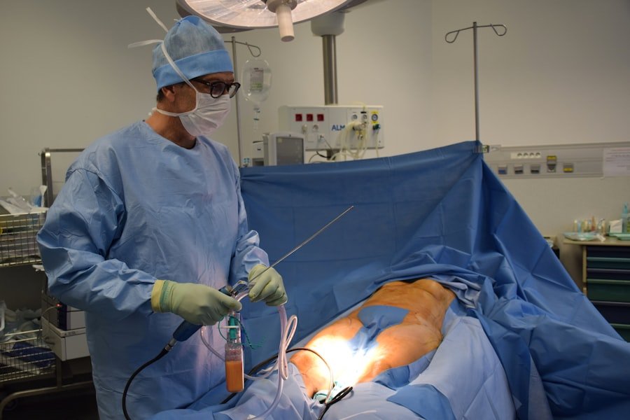
- The Operating Room Symphony: The Surgical Procedure Unfolds
This is where the meticulous planning translates into action. The operating room is a highly sterile, controlled environment where a team of dedicated professionals works in unison, guided by precision and expertise. Your LCA is here to orchestrate this complex ballet of instruments, technology, and skilled hands, explaining each critical phase of the acoustic neuroma surgery.
2.1. Anesthesia Induction: Entering a State of Rest
The journey into surgery begins with anesthesia. It’s designed to ensure you are completely unaware of the procedure and experience no pain.
2.1.1. Intravenous Access and Sedation
A nurse will place an intravenous (IV) line in your arm. This will be used to administer fluids, medications, and the anesthetic agents. You might receive some mild sedatives to help you relax before the general anesthesia is administered.
2.1.2. General Anesthesia Administration
The anesthesiologist will administer the anesthetic agents, typically through your IV, which will quickly render you unconscious. Breathing support will be provided through an endotracheal tube or a laryngeal mask airway.
2.1.3. Neuromonitoring Setup
While you are being prepared for surgery, specialized equipment will be set up to continuously monitor the function of your facial nerve and auditory pathways.
- Facial Nerve Monitoring (Electromyography – EMG): Small electrodes are placed on facial muscles to detect nerve activity.
- Auditory Brainstem Response (ABR) Monitoring: Electrodes are placed on your scalp to record electrical activity in the auditory pathways in response to sound stimuli. This helps the surgeon assess nerve function in real-time during the operation, crucial for hearing preservation efforts.
2.2. The Incision and Craniotomy: Accessing the Surgical Field
This is where the surgeon carefully creates an opening to reach the acoustic neuroma. The specific approach dictates the exact steps here.
2.2.1. Surgical Site Preparation and Sterilization
The skin area where the incision will be made is meticulously cleansed with antiseptic solutions to eliminate any potential for infection. Sterile drapes are then placed around the surgical field.
2.2.2. The Incision: A Carefully Placed Cut
- Retrosigmoid Approach: A curved incision is made behind the ear, extending from just above the ear towards the nape of the neck.
- Middle Fossa Approach: An incision is made above the ear and curves back.
- Translabyrintine Approach: An incision is made behind the ear, similar to the retrosigmoid, but the dissection proceeds through the mastoid bone.
2.2.3. The Craniotomy: Creating an Opening in the Skull
- Bone Flap Creation: Using a specialized surgical drill (craniotome), the neurosurgeon carefully removes a section of bone from the skull. The size and shape of this bone flap depend on the surgical approach and the size of the tumor.
- Dural Opening: Once the bone is removed, the dura mater – the tough outer membrane protecting the brain – is carefully incised and opened to expose the brain surface beneath.
2.3. Tumor Localization and Identification: Pinpointing the Target
With access secured, the surgeon now begins the delicate task of finding and isolating the acoustic neuroma.
2.3.1. Navigational Guidance and Intraoperative Ultrasound
Modern surgical suites often utilize neuronavigation systems. These systems use pre-operative MRI scans to create a 3D map of your brain, allowing the surgeon to precisely track their instruments in relation to anatomical structures, including the tumor. Intraoperative ultrasound may also be used to further delineate the tumor margins and critical structures.
2.3.2. Identification of the Internal Auditory Canal (IAC)
The surgeon will carefully identify the internal auditory canal (IAC), the bony channel through which the acoustic neuroma grows and where the facial and auditory nerves reside.
2.3.3. Dissection of Surrounding Structures: Preserving Vital Nerves
This is arguably the most critical and challenging part of the surgery. The surgeon meticulously dissects the tumor away from the vital nerves.
- Facial Nerve Identification and Preservation: Using microdissection techniques and often guided by intraoperative neuromonitoring, the surgeon carefully separates the facial nerve from the tumor. Preserving facial nerve function is a top priority.
- Cochlear Nerve Identification and Preservation (where possible): For tumors where hearing preservation is a goal, the surgeon will attempt to identify and preserve the cochlear nerve, though this can be very challenging with larger tumors.
2.4. Tumor Resection: The Delicate Removal Process
This is the heart of the operation, where the acoustic neuroma is carefully removed. The technique employed depends on the tumor’s size, consistency, and location.
2.4.1. Microsurgical Techniques and Instrumentation
All acoustic neuroma surgeries are performed using microsurgical techniques. This involves using high-powered microscopes with magnification up to 40x and specialized micro-instruments (tiny forceps, scissors, and dissectors) to work with extreme precision.
2.4.2. Tumor Debulking and Removal
- Ultrasonic Aspirator (CUSA): For larger tumors, an ultrasonic aspirator (CUSA) may be used. This device breaks up tumor tissue into small fragments with ultrasonic waves, which are then suctioned away, allowing for gradual removal without excessive manipulation of surrounding nerves.
- Endoscopic Assistance: In some cases, an endoscope – a small, flexible camera – may be used to provide improved visualization of difficult-to-reach areas or to confirm complete tumor removal.
- Gradual Resection: The tumor is typically removed in segments, working from the periphery inward, to minimize the risk of damaging crucial nerves.
2.4.3. Confirmation of Complete Tumor Removal
The surgeon will meticulously inspect the surgical bed to ensure that all visible tumor tissue has been removed. This is often aided by the microscopic view and intraoperative imaging. The goal is to achieve a gross total resection whenever possible, but this is balanced against the risk of functional deficits.
2.5. Reconstruction and Closure: Sealing the Surgical Field
Once the tumor is removed, the surgical site is prepared for closure, ensuring a secure and well-healed wound.
2.5.1. Hemostasis: Ensuring No Bleeding
The surgical team takes great care to meticulously control any bleeding points using cautery and hemostatic agents. This is crucial for preventing post-operative hematomas.
2.5.2. Dural Closure and Reconstruction
The dura mater is carefully repaired, often using sutures and sometimes with the aid of a dural graft material (e.g., pericranium, fascia lata, or synthetic materials) to create a watertight seal. This is vital to prevent cerebrospinal fluid (CSF) leaks.
2.5.3. Bone Graft Placement (if applicable)
If a significant bone flap was created, it will be repositioned and secured in place using tiny screws and plates to facilitate healing.
2.5.4. Layered Closure of Tissues and Skin
The layers of tissue above the bone are then meticulously closed with sutures. The skin incision is typically closed with sutures or surgical staples, often with an underlying drain to remove any excess fluid.
- The Post-operative Landscape: Navigating the Immediate Recovery
You’ve woken up. The surgery is complete, and you are now entering the crucial immediate post-operative period. This phase is about waking from anesthesia, managing initial pain and discomfort, and beginning the journey back to wellness. As your LCA, I’ll guide you through what to expect in the hours and days immediately following your surgery.
3.1. Awakening from Anesthesia: The Gentle Return to Consciousness
This is a gradual process, and it’s normal to feel disoriented as the anesthetic agents wear off.
3.1.1. Transfer to the Post-Anesthesia Care Unit (PACU)
You’ll be moved from the operating room to the PACU, also known as the recovery room. Here, highly trained nurses will closely monitor your vital signs (heart rate, blood pressure, oxygen saturation, and temperature).
3.1.2. Initial Assessment of Neurological Status
The medical team will perform initial checks of your consciousness, pupil response, and general neurological function. Any immediate concerns will be addressed promptly.
3.1.3. Pain and Nausea Management
Pain medication will be administered through your IV to manage discomfort from the surgical incision and the procedure itself. You may also receive medication to help with any nausea or vomiting that can occur after anesthesia.
3.2. In the Intensive Care Unit (ICU) or Step-Down Unit: Continuous Monitoring
Depending on the complexity of your surgery and your overall health, you may spend some time in the ICU or a specialized neuro-surgical step-down unit.
3.2.1. Enhanced Vital Sign and Neurological Monitoring
The level of monitoring will be intensified, with continuous observation of your vital parameters and neurological status. This allows for early detection of any potential complications.
3.2.2. Management of Intravenous Fluids and Medications
You’ll continue to receive IV fluids to maintain hydration and necessary medications, including antibiotics to prevent infection and pain relief.
3.2.3. Monitoring for Potential Complications
The team will be vigilant for signs of complications such as bleeding, infection, swelling, or cerebrospinal fluid (CSF) leaks.
3.3. Moving to a Regular Hospital Room: Beginning to Mobilize
As your condition stabilizes, you’ll be transferred to a regular hospital room, where the focus shifts to recovery and increasing independence.
3.3.1. Gradual Increase in Activity
- Early Mobilization: Nurses will encourage you to sit up in bed and, when you feel ready, to start taking short walks with assistance. This is crucial for preventing blood clots and promoting circulation.
- Assistance with Daily Activities: Initially, you may need help with tasks such as bathing, dressing, and eating. However, the goal is to gradually regain your independence.
3.3.2. Pain Management Transition: Oral Medications
Your pain management will transition from intravenous to oral medications as your comfort level improves.
3.3.3. Monitoring Incision Site and Drain Output
The surgical incision will be monitored for any signs of infection, redness, or swelling. If a drain is in place, its output will be carefully recorded.
3.4. Assessing Neurological Function: Evaluating Nerve Recovery
Regular assessments of your facial nerve, hearing, and balance will be conducted.
3.4.1. Facial Nerve Examinations
The medical team will repeatedly check for facial symmetry and movement, noting any improvements or areas of concern.
3.4.2. Hearing and Vestibular Assessments
While formal audiologist tests might be delayed, your ability to hear and your balance will be observed and inquired about. Any new or worsening symptoms will be documented.
3.4.3. Speech and Swallowing Evaluation
Depending on the extent of the surgery and potential nerve involvement, a speech therapist may assess your swallowing and speech to identify any difficulties and recommend interventions.
3.5. Dietary Adjustments and Hydration: Fueling Your Recovery
Your diet will be carefully managed to support healing and address any potential issues.
3.5.1. Starting with Liquids and Soft Foods
You’ll typically start with clear liquids and progress to soft foods as you tolerate them, ensuring you don’t strain your facial muscles or gag.
3.5.2. Ensuring Adequate Hydration
Staying well-hydrated is essential for overall recovery and to help prevent complications. You’ll be encouraged to drink plenty of fluids.
3.5.3. Managing Potential Swallowing Difficulties
If swallowing difficulties arise, your diet may be modified, and you might receive guidance from a speech therapist on techniques to improve swallowing safety.
- The Rehabilitation Ripple Effect: Regaining Function and Independence
Once you are discharged from the hospital, the journey of recovery truly unfolds in your own environment. This post-operative phase is dedicated to rebuilding strength, restoring function, and reclaiming your quality of life. As your LCA, I’ll map out the crucial elements of this rehabilitation process.
4.1. Discharge Planning: A Tailored Roadmap Home
Before you leave the hospital, a comprehensive discharge plan will be established, outlining everything you need for a successful transition.
4.1.1. Review of Discharge Instructions
You’ll receive detailed instructions covering medication schedules, wound care, activity restrictions, dietary recommendations, and warning signs to watch for. Make sure you understand every point and have a chance to ask questions.
4.1.2. Prescription for Medications and Therapies
Your prescriptions for pain relief, and any other necessary medications, will be provided. Referrals for outpatient therapies, such as physical therapy or occupational therapy, will also be made.
4.1.3. Scheduling Follow-Up Appointments
Key follow-up appointments with your surgeon, and potentially other specialists, will be scheduled. These are critical for monitoring your progress and addressing any emerging concerns.
4.2. Home Recovery and Wound Care: Nurturing the Healing Site
Your home becomes your primary recovery environment. Care for your surgical site is paramount.
4.2.1. Managing Surgical Incision and Drains
- Keeping the Area Clean and Dry: You’ll be instructed on how to gently clean around the incision and change dressings as needed. Keeping the wound clean and dry is crucial for preventing infection.
- Drain Management (if applicable): If you have a drain, you’ll be shown how to empty and care for it until it’s removed at a follow-up appointment.
4.2.2. Recognizing Signs of Infection
Be aware of the signs of infection, including increasing redness, swelling, warmth around the incision, fever, chills, or any discharge from the wound. Contact your doctor immediately if you notice any of these.
4.2.3. Activity Restrictions and Gradual Progression
Your surgeon will provide specific guidelines on physical activity. This typically involves starting with gentle movements and gradually increasing your range of motion and endurance as you heal. Avoid strenuous activities, heavy lifting, or anything that puts excessive strain on your head or incision for the initial period.
4.3. Physical Therapy: Reclaiming Balance and Mobility
Physical therapy is a cornerstone of recovery, especially for addressing issues related to balance, dizziness, and overall strength.
4.3.1. Vestibular Rehabilitation Therapy
This specialized therapy focuses on exercises designed to retrain your brain to compensate for impaired balance and vertigo. It can significantly improve your stability and reduce dizziness.
4.3.2. Strengthening and Endurance Exercises
Your physical therapist will guide you through exercises to rebuild muscle strength and improve your stamina, which may have been affected by the surgery and the neuroma itself.
4.3.3. Gait Training and Fall Prevention
Exercises will focus on improving your walking pattern (gait) and balance to reduce the risk of falls, a common concern after acoustic neuroma surgery.
4.4. Occupational Therapy: Adapting to Daily Life
Occupational therapy helps you regain independence in performing everyday activities.
4.4.1. Strategies for Daily Living
If you experience facial weakness, sensory changes, or difficulty with fine motor skills, an occupational therapist can teach you adaptive techniques and recommend assistive devices to help with tasks like eating, dressing, and grooming.
4.4.2. Home Modifications Recommendations
They can also assess your home environment and suggest modifications to make it safer and more accessible during your recovery.
4.4.3. Cognitive and Sensory Adaptation Strategies
If you experience any cognitive fogginess or sensory processing changes, an occupational therapist can provide strategies to help you adapt and manage these challenges.
4.5. Speech Therapy and Swallowing Support: Ensuring Safe and Effective Communication and Nutrition
If speech or swallowing issues arise, speech-language pathologists play a vital role.
4.5.1. Exercises for Speech Articulation and Resonance
If facial weakness affects your speech, therapy can focus on improving articulation, clarity, and voice quality.
4.5.2. Swallowing Exercises and Diet Modification
For dysphagia (difficulty swallowing), therapists provide exercises to strengthen swallowing muscles and may recommend texture-modified diets to ensure safe and efficient nutrition.
4.5.3. Addressing Facial Sensory Changes
Therapists can also help you manage sensory changes in your face, such as numbness or altered taste, by providing techniques for sensory re-education.
- Long-Term Outlook and Ongoing Management: Living Well Beyond Surgery
The surgery is a significant milestone, but it’s not the end of your journey. Your LCA is here to guide you through the long-term considerations and ongoing management that will ensure you live a full and healthy life post-acoustic neuroma surgery. This phase is about continued monitoring, adaptation, and embracing your new normal.
5.1. Follow-Up Medical Appointments: Continuous Monitoring and Evaluation
Regular check-ins with your medical team are crucial to track your recovery and detect any potential late-onset issues.
5.1.1. Neurological Assessments
Your surgeon will continue to assess your neurological function, including facial nerve strength, hearing, and balance. These assessments help gauge nerve recovery and identify any areas requiring further attention.
5.1.2. Hearing and Vestibular Evaluations
Periodic audiology and vestibular function tests will be performed to monitor your hearing and balance, particularly if hearing preservation was a goal. This helps track any changes and adjust management strategies as needed.
5.1.3. Imaging for Recurrence Surveillance
While recurrences are rare after complete resection, follow-up MRI scans are typically recommended for a period to ensure no regrowth of the tumor occurs. The frequency and duration of these scans will be determined by your surgeon.
5.2. Managing Residual Symptoms and Functional Changes: Adapting and Thriving
It’s common for some residual symptoms or functional changes to persist. The focus here is on adaptation and finding strategies to manage them effectively.
5.2.1. Ongoing Management of Facial Weakness
If facial weakness persists, continued work with physical or occupational therapists, or even exploring surgical options for facial reanimation, might be considered. Medical and cosmetic camouflage techniques can also be helpful.
5.2.2. Strategies for Hearing Loss and Tinnitus
- Hearing Aids and Assistive Listening Devices: For hearing loss, hearing aids can be very beneficial. Assistive listening devices, such as special telephones or classroom amplification systems, can further enhance your ability to communicate.
- Tinnitus Management: Strategies for managing tinnitus may include sound therapy, cognitive behavioral therapy (CBT), and relaxation techniques to reduce its intrusiveness.
5.2.3. Coping with Balance and Dizziness Issues
Continued engagement in vestibular rehabilitation exercises, mindfulness, and lifestyle adjustments can help manage persistent balance or dizziness challenges. Avoiding triggers and ensuring a safe environment are key.
5.3. Emotional and Psychological Well-being: The Importance of Mental Health
Undergoing surgery and navigating a lengthy recovery can take a significant emotional toll. Prioritizing your mental health is as important as your physical recovery.
5.3.1. Addressing Anxiety and Depression
It’s normal to experience fluctuations in mood. If you are struggling with anxiety or depression, don’t hesitate to seek professional help. Talking with a therapist or counselor can provide invaluable support.
5.3.2. Connecting with Support Groups
Connecting with others who have undergone similar experiences can be incredibly empowering. Support groups offer a safe space to share concerns, exchange coping strategies, and find encouragement.
5.3.3. Maintaining Social Connections and Engaging in Hobbies
Staying connected with friends and family and engaging in activities you enjoy can significantly boost your morale and overall well-being. Pace yourself and adapt activities as needed.
5.4. Lifestyle Modifications for Long-Term Health: Embracing a Healthy Future
Adopting healthy lifestyle habits can help optimize your recovery and promote overall well-being.
5.4.1. Balanced Nutrition and Hydration
Continue to prioritize a nutritious diet rich in fruits, vegetables, and lean proteins to support continued healing and energy levels. Staying well-hydrated remains important.
5.4.2. Regular Exercise and Physical Activity
Incorporate regular, moderate exercise into your routine as recommended by your doctor. This helps maintain strength, cardiovascular health, and a positive mood.
5.4.3. Stress Management Techniques
Continue to practice stress-reducing techniques such as mindfulness, meditation, yoga, or deep breathing exercises. Managing stress is beneficial for both physical and mental health.
5.5. Patient Advocacy and Empowerment: Empowering Your Health Journey
You are the central figure in your health journey. Understanding your condition and actively participating in your care is key to achieving the best possible outcomes.
5.5.1. Staying Informed About Your Condition
Continue to educate yourself about acoustic neuromas and your specific situation. Knowledge empowers you to have more informed discussions with your medical team.
5.5.2. Communicating Openly with Your Healthcare Team
Maintain open and honest communication with your surgeon, therapists, and other healthcare providers. Report any new symptoms, concerns, or changes in your condition promptly.
5.5.3. Setting Realistic Goals and Celebrating Milestones
Recovery is a marathon, not a sprint. Set realistic goals for yourself, celebrate the milestones you achieve along the way, and be patient and compassionate with yourself throughout the process. Your resilience is your greatest asset.
FAQs
What is an acoustic neuroma?
An acoustic neuroma is a non-cancerous tumor that develops on the main nerve leading from the inner ear to the brain. It can cause hearing loss, ringing in the ear, and unsteadiness.
When is surgery recommended for acoustic neuroma?
Surgery is typically recommended for acoustic neuroma if the tumor is large, growing rapidly, or causing significant symptoms such as hearing loss, balance problems, or facial numbness.
What are the steps involved in acoustic neuroma surgery?
Acoustic neuroma surgery involves several steps, including making an incision behind the ear, removing a portion of the skull bone, identifying and carefully separating the tumor from the surrounding nerves, and closing the incision.
What are the potential risks and complications of acoustic neuroma surgery?
Potential risks and complications of acoustic neuroma surgery include hearing loss, facial weakness, balance problems, cerebrospinal fluid leakage, and infection.
What is the recovery process like after acoustic neuroma surgery?
Recovery after acoustic neuroma surgery can vary, but typically involves a hospital stay of several days, followed by a period of rest and rehabilitation. Patients may experience temporary facial weakness, balance issues, and hearing changes. Full recovery can take several weeks to months.