As the Listicle Content Architect, you’ve been tasked with crafting the definitive guide to facial nerve complications. You know that clarity, accessibility, and thoroughness are key. Your goal is to make complex medical information digestible for a broad audience, empowering them with knowledge about the facial nerve and its potential pitfalls. You’ll weave in detailed explanations, practical advice, and re-assuring insights, all presented in your signature engaging and authoritative tone.
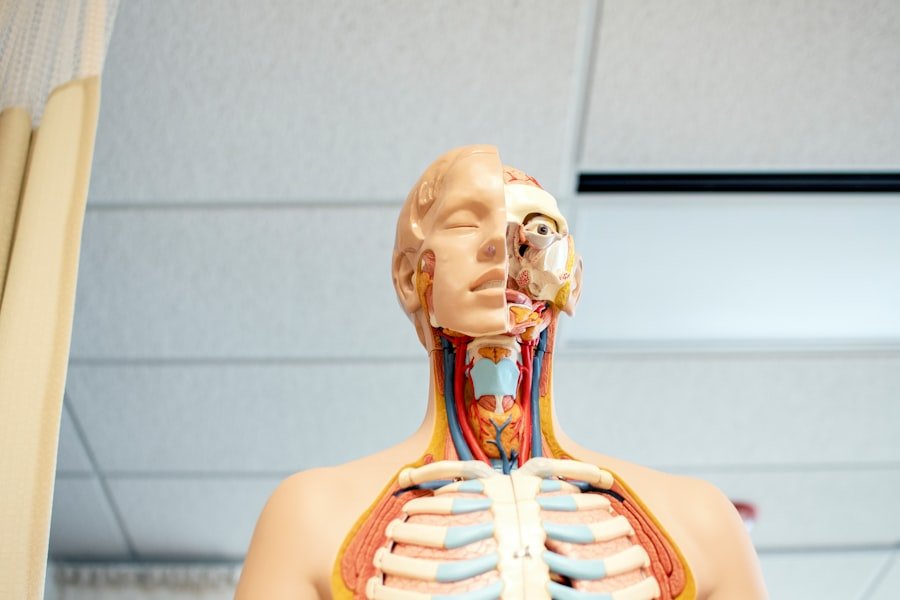
Before diving into the complications, it’s crucial to understand the star of the show: your facial nerve, also known as the seventh cranial nerve (CN VII). Think of it as the conductor of your face’s symphony of expressions and sensations. It’s a remarkably complex nerve, originating deep within your brainstem and embarking on a long, intricate journey through your skull and into your face. Its multifunctionality is astounding; it’s responsible for everything from your subtlest smile to the protective blink of your eye, and even plays a role in taste and tear production.
The Brainstem Origin: Where the Symphony Begins
Your facial nerve’s journey starts in the pons, a part of your brainstem. Here, specialized neurons form the nucleus of the facial nerve. This is where all the commands for facial movement originate. Imagine this as the central control room, where instructions are meticulously crafted before being sent out to the muscles. The nerve then winds its way through the internal auditory canal, a narrow passage in your temporal bone, before emerging from the skull at the stylomastoid foramen, a small opening just below your ear.
The Intricate Pathway Through the Temporal Bone
The portion of the facial nerve that travels through the temporal bone is particularly vulnerable due to the tight confines of this bony canal. This segment is also where the nerve gives off crucial branches. Just before it exits the skull, the facial nerve branches off a nerve called the chorda tympani, which plays a vital role in transmitting taste sensation from the front two-thirds of your tongue. It also gives off a branch that stimulates salivary glands, contributing to saliva production – essential for digestion and keeping your mouth moist. This intricate pathway highlights why even subtle pressure or inflammation in this area can have significant consequences.
The Branching Network: Controlling Your Expressions
Once the facial nerve exits the stylomastoid foramen, it fans out into a complex network of branches that innervate virtually every muscle involved in facial expression. You have muscles for raising your eyebrows, wrinkling your nose, puffing out your cheeks, smiling, frowning, and closing your eyes. Each of these actions is a precisely coordinated effort orchestrated by the facial nerve’s commands. These branches are specific, meaning that damage to one branch might affect a particular area of the face while leaving others intact, leading to localized weakness or paralysis.
Sensory and Autonomic Functions: Beyond Movement
While the motor functions of the facial nerve are most apparent, it’s also a sensory and autonomic powerhouse. You’ve already learned about the chorda tympani‘s role in taste sensation. Additionally, the facial nerve carries sensory information from a small area around your ear. On the autonomic side, it stimulates the lacrimal glands, responsible for tear production, and the submandibular and sublingual salivary glands, contributing to saliva. This explains why some facial nerve issues can also manifest as dry eyes or a dry mouth, or conversely, excessive tearing or salivation.
Why Understanding This Matters for Complications
By appreciating the facial nerve’s intricate anatomy and its diverse functions, you can better grasp why complications arise. Any disruption along its long pathway – from the brainstem to the tiniest muscle fibers – can lead to a cascade of symptoms. Whether it’s direct trauma, inflammation, viral infections, or other underlying conditions, understanding the nerve’s structure helps pinpoint the likely cause and the potential consequences.
Common Causes of Facial Nerve Complications
The facial nerve, despite its resilience, is susceptible to a variety of insults. These can range from sudden, acute events to gradual, chronic processes. Recognizing the potential culprits is the first step in understanding and managing facial nerve complications. Your mission as the LCA is to demystify these causes, making them relatable and understandable to your audience.
Bell’s Palsy: The Most Frequent Culprit
By far the most common cause of sudden unilateral facial paralysis is Bell’s palsy. You likely know someone who has experienced it. It’s often characterized by a sudden onset of weakness or complete paralysis on one side of the face, typically developing over a few hours to a few days. While the exact cause remains unknown, it’s widely believed to be related to inflammation and swelling of the facial nerve, likely triggered by a viral infection, such as the herpes simplex virus (HSV). This inflammation compresses the nerve within its bony canal, disrupting its ability to send signals to the facial muscles.
Other Viral and Bacterial Infections: A Wider Net
While Bell’s palsy is the most common, other viral and bacterial infections can also wreak havoc on the facial nerve. Shingles (herpes zoster), caused by the varicella-zoster virus (the same virus that causes chickenpox), can affect the facial nerve, leading to a condition known as Ramsay Hunt syndrome. This syndrome is characterized by a painful rash in and around the ear, facial paralysis, and sometimes hearing loss or vertigo. Other infections, such as Lyme disease (a bacterial infection transmitted by ticks), can also indirectly affect the facial nerve through inflammation or damage to surrounding tissues.
Traumatic Injuries: Direct Hits and Surgical Risks
The facial nerve’s journey makes it vulnerable to trauma. This can include direct blows to the head or face, fractures of the temporal bone (the bone surrounding the ear), or injuries sustained during surgical procedures. For instance, surgery involving the ear, parotid gland (a large salivary gland in the face), or brainstem can inadvertently damage the facial nerve. While surgeons take great care to avoid this, it remains a risk, and understanding this possibility is important for patients undergoing such procedures. The severity of the complication depends on the extent of the damage – a minor stretch might lead to temporary weakness, while a complete transection (severing) could result in permanent paralysis.
Tumors and Cysts: Pressure and Intrusion
Tumors and cysts that grow along the pathway of the facial nerve can cause progressive damage by directly compressing or invading the nerve. These can occur at various points, including within the brainstem, along the nerve’s course through the temporal bone, or near the parotid gland where the nerve branches extensively. The symptoms often develop gradually as the tumor grows, leading to increasing weakness or paralysis. Early detection and treatment of these underlying growths are crucial to minimize nerve damage.
Autoimmune and Inflammatory Conditions: The Body’s Own Attack
Less commonly, autoimmune and inflammatory conditions can target the facial nerve. Diseases like sarcoidosis, which can cause inflammation in various organs, can sometimes affect the nerves, including the facial nerve. Similarly, conditions that cause widespread inflammation can lead to facial nerve dysfunction. Your LCA mission here is to convey that sometimes the body’s own immune system can be the cause of the problem, leading to intricate and challenging cases.
Recognizing the Symptoms: What to Look For

The symptoms of facial nerve complications can be varied and depend on which part of the nerve is affected and the extent of the damage. Crucially, the onset can be sudden or gradual, adding to the anxiety patients might feel. Your role is to equip your audience with the knowledge to identify these signs so they can seek appropriate medical attention promptly.
Sudden Onset of Facial Weakness or Paralysis
The hallmark symptom of many facial nerve complications, particularly Bell’s palsy, is the sudden onset of weakness or complete paralysis on one side of the face. This often develops over a period of hours to a few days. You might notice that one side of your face droops, making it difficult to smile, frown, or close your eye on that side. The affected side may feel “numb” or “heavy,” though true sensation is usually preserved. This unilateral nature is a key indicator, differentiating it from stroke, which often affects both sides of the face.
Difficulty with Facial Expressions
As a direct result of muscle weakness, you’ll experience difficulty with various facial expressions. Your smile might be lopsided, your forehead may not wrinkle on the affected side when you try to raise your eyebrows, and you might find it hard to squint or close your eye tightly. Puffed-out cheeks might deflate unevenly, and you might have trouble pursing your lips. These are visible, often distressing, manifestations of the nerve’s compromised signaling.
Eyelid and Eye Issues: The Vulnerable Eye
The facial nerve plays a critical role in controlling the muscles that close your eyelid. When this function is impaired, you may have trouble closing your eye completely on the affected side. This can lead to dryness, irritation, and increased vulnerability to injury for the eye. Tears might also be affected, leading to either excessive tearing (if the nerve’s ability to drain tears is compromised) or reduced tear production (if the lacrimal gland stimulation is impaired). Corneal abrasions are a significant concern due to the inability to protect the eye.
Alterations in Taste and Salivation
As mentioned earlier, the facial nerve carries taste signals and influences saliva production. Therefore, complications affecting certain parts of the nerve can lead to altered taste sensation, particularly on the front two-thirds of your tongue. You might perceive tastes as dulled, metallic, or absent altogether. Similarly, you might notice changes in salivation, such as a dry mouth or, less commonly, an excessive flow of saliva on the affected side.
Sound Sensitivity (Hyperacusis)
In some cases, damage to the facial nerve can affect a tiny muscle in the middle ear called the stapedius muscle. This muscle helps to dampen loud sounds. When the nerve controlling it is compromised, you might experience hyperacusis, which is an increased sensitivity to everyday sounds. Sounds that were once tolerable may become uncomfortably loud or even painful. This can be a particularly unsettling symptom for those affected.
Diagnosis and Assessment: Pinpointing the Problem
| Complication | Description |
|---|---|
| Facial Nerve Paralysis | Loss of movement in the muscles of the face due to damage to the facial nerve. |
| Facial Muscle Weakness | Reduced strength in the facial muscles, leading to difficulty in making facial expressions. |
| Facial Nerve Palsy | Weakness or paralysis of the facial muscles due to nerve damage. |
| Facial Numbness | Lack of sensation in the face, often accompanied by tingling or loss of feeling. |
When you suspect a facial nerve complication, seeking prompt medical evaluation is essential. A thorough diagnosis involves a combination of medical history, physical examination, and sometimes specialized tests to accurately identify the cause and extent of the nerve dysfunction. Your expertise as an LCA means you’ll guide your reader through this process with confidence and clarity.
The Neurological Examination: A Deep Dive
The cornerstone of diagnosing facial nerve complications is the neurological examination. Your doctor will meticulously assess the strength and symmetry of facial movements on both sides of your face. They will ask you to perform a series of actions: raising your eyebrows, squeezing your eyes shut, puffing out your cheeks, smiling, and frowning. They will observe the range and quality of these movements, noting any asymmetry or weakness. This detailed assessment helps localize the problem to the facial nerve and provides clues about the severity of the damage.
Gathering Your Medical History: The Narrative of Your Health
Your medical history is a crucial piece of the diagnostic puzzle. Your doctor will inquire about the onset and progression of your symptoms, any recent illnesses or infections you’ve experienced (such as a cold or flu), any previous episodes of facial weakness, your medical history (including conditions like diabetes or hypertension), and any significant life events or stressors. Information about any recent surgeries or injuries to the head or face is also vital. This narrative helps identify potential underlying causes and risk factors.
Ruling Out Stroke: A Critical Distinction
It’s imperative to rule out a stroke as the cause of facial weakness, especially if the onset is very sudden and accompanied by other neurological symptoms like weakness in an arm or leg, difficulty speaking, or disorientation. While both stroke and facial nerve complications can cause facial drooping, a stroke typically affects a broader area of the brain and often involves more generalized neurological deficits. Emergency medical services are trained to quickly differentiate between these conditions, and time is of the essence in stroke management.
Imaging Studies: A Visual Roadmap
In certain situations, imaging studies may be recommended to gain a clearer picture of the facial nerve and surrounding structures. This can include:
- MRI (Magnetic Resonance Imaging): This is often the preferred imaging technique as it provides detailed images of soft tissues, including nerves and the brain. It can help identify inflammation, tumors, or other abnormalities affecting the facial nerve or its pathways.
- CT (Computed Tomography) Scan: A CT scan is useful for visualizing bony structures and can help identify fractures of the temporal bone or other bone abnormalities that might be compressing the facial nerve. It’s also often used in emergency settings to quickly assess for stroke.
These imaging modalities provide invaluable visual information for diagnosis and treatment planning.
Electrophysiological Tests: Measuring Nerve Function
Electrophysiological tests can be used to assess the electrical activity of the facial nerve and its muscles. These tests can help determine the degree of nerve damage and predict the likelihood of recovery. The most common tests include:
- EMG (Electromyography): This test measures the electrical activity in muscles. Needles are inserted into specific facial muscles to record their electrical signals, both at rest and during voluntary movement. This helps assess muscle health and identify any abnormalities caused by nerve damage.
- Nerve Conduction Studies (NCS): In NCS, a mild electrical impulse is applied to the nerve to measure how quickly and strongly it conducts signals. This can help quantify the severity of nerve damage and distinguish between nerve damage and muscle problems.
These tests provide objective measures of nerve function, complementing the clinical assessment.
Treatment and Management Strategies: Restoring Function and Reducing Discomfort
The approach to treating facial nerve complications is tailored to the underlying cause and the severity of the nerve damage. Your LCA goal is to provide a comprehensive overview of the various therapeutic avenues available, empowering individuals with knowledge about their recovery journey.
Medications: Addressing Inflammation and Infection
The primary goal of medication in many facial nerve complications, particularly Bell’s palsy, is to reduce inflammation and combat any underlying infection.
- Corticosteroids: Medications like prednisone are often prescribed to reduce inflammation and swelling of the facial nerve. Early administration of corticosteroids, ideally within 72 hours of symptom onset, can significantly improve the chances of recovery.
- Antiviral Medications: If a viral infection, such as herpes simplex or herpes zoster, is suspected or confirmed, antiviral medications (e.g., acyclovir, valacyclovir) may be prescribed. These medications can help suppress viral activity and may be used in conjunction with corticosteroids, especially in cases of Ramsay Hunt syndrome.
Physical Therapy and Facial Rehabilitation: Re-educating Your Muscles
Physical therapy and facial rehabilitation are crucial components of recovery. A skilled therapist will guide you through exercises designed to:
- Maintain muscle tone: Gentle exercises can help prevent muscles from atrophying (wasting away) due to disuse.
- Improve facial muscle strength and coordination: As nerve function begins to return, targeted exercises help retrain the brain-muscle connection, improving voluntary control and symmetry.
- Prevent contractures: Without proper movement, facial muscles can become stiff and develop contractures. Therapy helps prevent this.
- Teach compensatory strategies: Therapists can teach you techniques to improve your appearance and function, such as methods for eating, speaking, and protecting your eye.
Consistency is key with this modality; your dedication to the prescribed exercises will directly impact your recovery outcome.
Eye Care: Protecting Your Most Vulnerable Sense
Given the potential for eyelid dysfunction, meticulous eye care is paramount. This may include:
- Lubricating eye drops: Artificial tears help keep the eye moist and prevent dryness and irritation.
- Eye ointments: These can provide longer-lasting lubrication, especially at night.
- Taping the eyelid shut: In severe cases, taping the eyelid closed at night is essential to protect the cornea from damage.
- Eye patches: These can provide protection during the day and help reduce light sensitivity.
Your ophthalmologist will work closely with your neurologist to ensure optimal eye health throughout your recovery.
Surgical Interventions: When Other Options Aren’t Enough
In some cases, surgical interventions may be considered, though they are not typically the first line of treatment for conditions like Bell’s palsy.
- Surgical Decompression: In the past, surgical decompression of the facial nerve within its bony canal was sometimes performed to relieve pressure on the nerve. However, current evidence suggests that this procedure may not significantly improve outcomes for Bell’s palsy and carries its own risks. It might be considered in specific, carefully selected cases if there’s strong evidence of nerve compression.
- Nerve Grafts or Reconstructive Surgery: For individuals with severe nerve damage due to trauma or tumor removal, nerve grafting or other reconstructive surgeries may be an option. These procedures aim to bridge gaps in the damaged nerve or reroute nearby healthy nerves to restore function to paralyzed facial muscles. These are complex procedures requiring specialized surgical expertise.
Your LCA knowledge ensures you highlight that surgery is usually a last resort or for specific trauma-related scenarios.
Botox Injections and Other Emerging Therapies
As the field of facial nerve rehabilitation evolves, new and innovative therapies are emerging. Botulinum toxin (Botox) injections, for instance, can be used strategically to reduce muscle overactivity or spasms in facial muscles and to help improve facial symmetry in some cases. Researchers are also exploring the potential of stem cell therapy and other regenerative approaches for nerve repair, offering hope for more comprehensive recovery in the future. Your role is to keep your audience informed about the frontiers of treatment.
Prognosis and Long-Term Outlook: Navigating Your Recovery Journey
Understanding your prognosis and the potential long-term outlook is vital for managing expectations and maintaining motivation throughout the recovery process. As the Listicle Content Architect, you’ll present this information with empathy and an emphasis on the many factors that influence the journey.
Factors Influencing Recovery: A Multifaceted Picture
The prognosis for recovery from facial nerve complications is highly variable and depends on several key factors:
- Cause of the Complication: Conditions like Bell’s palsy generally have a good prognosis, with most individuals making a significant recovery. However, complications arising from stroke, trauma, or tumors may have a less favorable outlook.
- Severity of Nerve Damage: Mild injuries that cause temporary inflammation or compression tend to recover more readily than complete nerve transections or severe structural damage.
- Timeliness of Treatment: Prompt diagnosis and initiation of appropriate treatment, particularly corticosteroids for Bell’s palsy, can significantly improve the chances and speed of recovery.
- Patient’s Overall Health: Age, underlying medical conditions (such as diabetes), and the individual’s general health status can influence their body’s ability to heal and regenerate.
- Adherence to Rehabilitation: Diligent participation in physical therapy and facial exercises is crucial for maximizing functional recovery and regaining muscle control.
Your LCA skill is to convey that recovery is not a one-size-fits-all scenario.
Typical Recovery Timeline: Patience and Progress
For Bell’s palsy, the typical recovery timeline can vary, but many individuals begin to see improvements within two to three weeks after symptom onset. Full recovery, however, can take anywhere from three months to a year. It’s important to remember that recovery is often a gradual process, with noticeable improvements occurring incrementally. Some individuals may experience a complete return of facial function, while others might have lingering mild weakness or asymmetry.
Potential for Residual Effects: Understanding Lingering Challenges
While many individuals recover fully, some may experience residual effects even after completing their treatment and rehabilitation. These can include:
- Synkinesis: This is a common long-term effect where voluntary movement of one facial muscle triggers an involuntary movement of another. For example, smiling might cause the eyelid on the same side to partially close.
- Persistent Weakness or Asymmetry: Some individuals may have mild, persistent weakness or asymmetry in certain areas of the face.
- Facial Spasms (Hemifacial Spasm): In some cases, facial nerve damage can lead to involuntary twitching or spasms of facial muscles.
- Altered Sensation: While rare, some individuals might experience persistent changes in taste or sensation around the ear.
Your LCA duty is to prepare your audience for these possibilities without causing undue alarm.
The Importance of Continued Monitoring and Support
Even after apparent recovery, continued monitoring and ongoing support can be beneficial. Regular follow-up appointments with your healthcare provider allow for assessment of any lingering issues and adjustment of rehabilitation strategies as needed. Connecting with support groups or patient advocacy organizations can provide valuable emotional support, shared experiences, and practical advice from others who have navigated similar challenges. Your aim is to empower individuals to be active participants in their ongoing well-being.
FAQs
What are the common causes of facial nerve complications?
Facial nerve complications can be caused by a variety of factors, including viral infections (such as Bell’s palsy), trauma or injury to the face or head, tumors, and certain medical conditions such as diabetes and high blood pressure.
What are the symptoms of facial nerve complications?
Symptoms of facial nerve complications can include facial weakness or paralysis, twitching or spasms in the face, drooping of the eyelid or corner of the mouth, loss of taste, and difficulty closing the eye on the affected side.
How are facial nerve complications diagnosed?
Facial nerve complications are typically diagnosed through a physical examination, including tests of facial muscle strength and movement. Additional tests such as imaging studies (MRI or CT scan) and nerve conduction studies may also be used to determine the cause and extent of the complication.
What are the treatment options for facial nerve complications?
Treatment for facial nerve complications depends on the underlying cause and severity of the condition. Options may include medication (such as corticosteroids or antiviral drugs), physical therapy, surgery, and in some cases, supportive measures such as eye protection and facial exercises.
What is the prognosis for facial nerve complications?
The prognosis for facial nerve complications varies depending on the cause and extent of the condition. In many cases, the majority of patients experience significant improvement in symptoms over time, while others may require ongoing management to address long-term effects.