
- Understanding the Role of Hearing Tests in Acoustic Neuroma Detection
You’re likely here because you, or someone you know, has been experiencing concerning changes in hearing – perhaps a persistent ringing, a feeling of fullness in one ear, or a noticeable decline in sound clarity. If this sounds familiar, you’re in the right place. As the Listicle Content Architect, I specialize in demystifying complex health topics, and today, we’re diving deep into the crucial role of hearing tests when an acoustic neuroma is suspected.
What is an Acoustic Neuroma?
Before we explore the tests, it’s essential to understand what an acoustic neuroma is. You see, it’s a non-cancerous tumor that develops on the vestibulocochlear nerve, which connects your inner ear to your brain. This nerve is responsible for two vital functions: hearing and balance. Because the tumor grows slowly and typically on just one side, its effects can be subtle at first, often leading to a gradual onset of symptoms that might be initially dismissed or attributed to other common ear issues.
Why Are Hearing Tests So Important?
The acoustic neuroma’s location on the vestibulocochlear nerve makes hearing issues one of its most prominent early signs. Therefore, hearing tests aren’t just a routine check-up in this context; they are diagnostic powerhouses. They act as your first line of defense, providing objective data that can point towards the presence of a tumor. Without these specialized evaluations, detecting an acoustic neuroma in its early, most treatable stages can be incredibly challenging.
The Subtle Nature of Early Symptoms
You might be wondering why you haven’t noticed more dramatic symptoms. Acoustic neuromas often grow at a snail’s pace. The pressure they exert on the delicate nerve fibers of the vestibulocochlear nerve can initially manifest as a gradual loss of high-frequency hearing, or a persistent ringing that you can’t ignore. These are the kinds of symptoms that might lead you to wonder, “Is my hearing changing?” and prompt you to seek professional advice.
Differentiating from Other Hearing Problems
It’s also vital to recognize that not all hearing changes are due to acoustic neuromas. Many everyday factors, like exposure to loud noise, aging, or fluid in the middle ear, can affect your hearing. This is precisely where the precision of hearing tests comes into play. They are designed to differentiate between the types of hearing loss and identify patterns that are more characteristic of a vestibular schwannoma (the medical term for acoustic neuroma).
The Goal: Early Detection and Intervention
Our ultimate goal here is early detection. The earlier an acoustic neuroma is identified, the greater the likelihood of successful treatment with minimal impact on your hearing and balance. Hearing tests are instrumental in achieving this goal by providing the foundational evidence that might lead to further, more specific diagnostic imaging. You’re not just getting your hearing measured; you’re actively participating in a process that safeguards your neurological health.
- Audiological Evaluations: The Cornerstone of Acoustic Neuroma Hearing Assessment
When you visit an audiologist because of concerns about hearing loss or other ear-related symptoms suggestive of an acoustic neuroma, you’ll undergo a series of detailed audiological evaluations. These tests are meticulously designed to assess your hearing across different frequencies and sound intensities, as well as to probe the intricate workings of your auditory system. You’re not just going to hear a beep; you’re participating in a comprehensive investigation.
Pure-Tone Audiometry: Mapping Your Hearing Thresholds
This is often the first test you’ll experience, and it’s fundamental. You’ll be seated in a soundproof booth, given a button to press whenever you hear a tone. The audiologist will present you with sounds of varying pitches (frequencies) and loudness (intensities) to both ears. They’ll be looking for your “hearing threshold” – the softest sound you can hear at each frequency.
What the Audiologist is Looking For:
- Type of Hearing Loss: Is it conductive (problems in the outer or middle ear), sensorineural (problems in the inner ear or nerve), or a combination? Acoustic neuromas typically cause sensorineural hearing loss.
- Degree of Hearing Loss: How significant is the loss? Mild, moderate, severe, or profound?
- Configuration of Hearing Loss: Does the loss affect specific frequencies more than others? A characteristic pattern for acoustic neuroma is a gradual decline in high-frequency hearing, often asymmetrical (worse in one ear than the other). This asymmetry is a key indicator that prompts further investigation.
Speech Audiometry: How Well Do You Understand Speech?
Hearing pure tones is one thing, but understanding speech in a noisy environment is another. Speech audiometry assesses your ability to hear and comprehend spoken words. You’ll typically hear spoken words at different loudness levels and be asked to repeat them.
Key Components of Speech Audiometry:
- Speech Reception Threshold (SRT): This is the softest level at which you can correctly repeat 50% of spoken words. It’s usually measured using simple two-syllable words. The SRT should align closely with your pure-tone thresholds, providing a cross-check for accuracy.
- Word Recognition Score (WRS) or Speech Discrimination Score (SDS): This test is crucial for acoustic neuroma assessment. You’ll hear lists of single-syllable words at a comfortable listening level, and your task is to repeat them. The percentage of words you identify correctly is your WRS. A significant WRS deficit in one ear, disproportionate to the pure-tone hearing loss at those frequencies, is a strong red flag for acoustic neuroma. This is because the tumor can distort the auditory signal before it even reaches the brain, impacting clarity.
Tympanometry: Examining Middle Ear Function
While acoustic neuromas primarily affect the inner ear and nerve, a complete audiological workup often includes tympanometry. This non-invasive test measures the movement of your eardrum and assesses the pressure in your middle ear.
What Tympanometry Reveals:
- Middle Ear Health: It helps rule out common causes of conductive hearing loss like earwax blockage, fluid in the middle ear (otitis media), or eardrum perforations.
- Reflexes: Acoustic reflexes are involuntary contractions of small muscles in the middle ear that occur in response to loud sounds. They are tested to assess the functionality of the auditory pathway from the ear to the brainstem. In acoustic neuroma cases, you might see abnormal or absent acoustic reflexes, especially at lower sound levels, because the tumor can interfere with the signal transmission through the nerve.
Otoacoustic Emissions (OAEs): A Quick Check of Inner Ear Function
Otoacoustic emissions are faint sounds produced by the delicate hair cells in your cochlea (the organ of hearing in the inner ear). These sounds are generated in response to external sound stimuli. OAEs are objectively measured using a small probe placed in your ear canal.
The Significance of OAEs:
- Cochlear Health: The presence of OAEs indicates that your outer hair cells in the cochlea are functioning properly. If OAEs are absent or significantly reduced in one ear, it strongly suggests a problem within the cochlea or along the auditory nerve.
- Sensitivity to Early Damage: OAEs are particularly sensitive to damage to the outer hair cells, which can be an early consequence of acoustic neuroma pressure. Even if you can still hear tones at higher volumes, a lack of OAEs can be a compelling clue for further investigation.
- Specialized Hearing Tests for Acoustic Neuroma Suspicions
When the initial audiological evaluations reveal anomalies – particularly asymmetrical hearing loss, poor speech discrimination out of proportion to pure-tone loss, or absent acoustic reflexes – it’s time to move on to more specialized tests. These are designed to more precisely pinpoint the nature of the problem and provide stronger evidence for or against an acoustic neuroma. You’re moving from a broad overview to a focused examination.
Acoustic Reflex Decay Testing: A Sensitive Indicator
This test is a more in-depth look at the acoustic reflex. After a loud, sustained tone is presented to elicit the acoustic reflex, the audiologist will monitor how long that reflex is maintained while the tone continues.
What Acoustic Reflex Decay Tells You:
- Nerve Integrity: A normal acoustic reflex should persist for as long as the stimulus is present. If the reflex “decays” or fades away rapidly (typically within the first few seconds of the sustained tone), it strongly suggests a problem with the auditory nerve. This is because the nerve fibers, compromised by the pressure of a growing tumor, can’t sustain their signal transmission.
- High Sensitivity: Acoustic reflex decay testing is considered one of the most sensitive audiological tests for detecting acoustic neuromas, even when the pure-tone hearing loss is relatively mild. You might hear the tone, but the nerve’s ability to “hold” that signal under sustained stimulus is compromised.
Auditory Brainstem Response (ABR) Testing: Mapping the Neural Pathway
The Auditory Brainstem Response (ABR) test, sometimes called the Brainstem Evoked Response Audiometry (BERA), is a highly objective diagnostic tool. It measures the electrical activity that travels from your ear through the auditory nerve to your brainstem in response to sound. You don’t need to actively participate; it’s all about measuring your body’s involuntary responses.
How ABR Works and What It Shows:
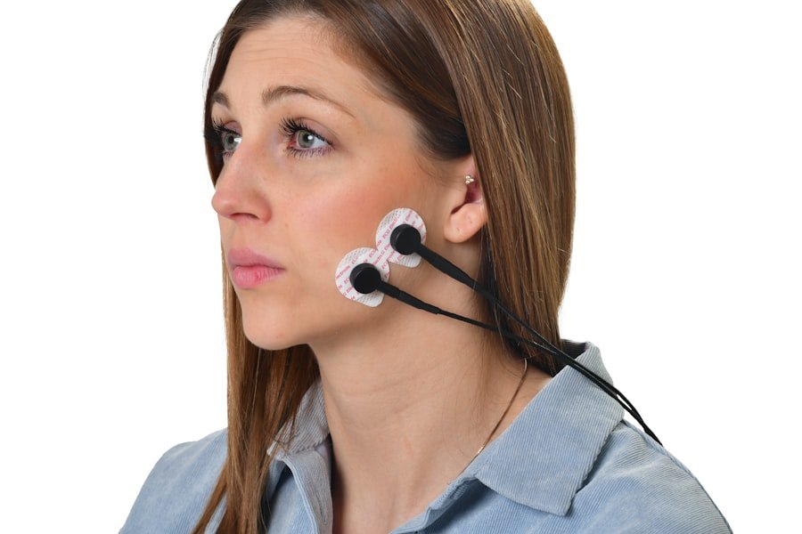
- Electrodes and Sound Stimuli: Small electrodes are placed on your scalp and earlobes. You’ll then hear a series of clicking sounds through earphones. The electrodes record the faint electrical signals generated by your auditory nerve and brainstem pathways as they process these sounds.
- Waveforms and Latencies: The ABR test generates a series of waveforms, each representing a specific point in the auditory pathway. Key metrics include the latency (the time it takes for each wave to appear) and the amplitude (the strength of the signal).
- Acoustic Neuroma Indicators: An acoustic neuroma can delay the transmission of signals along the auditory nerve. This will manifest as prolonged latencies for specific waves (particularly Wave V, which originates in the brainstem). The audiologist will compare the latencies between your ears. A significant difference in latency between the left and right ear, or abnormally long latencies in one ear, is a strong indicator of a potential acoustic neuroma. It essentially shows a “traffic jam” on the nerve.
Binaural Speech Testing: Assessing Listening in Two Ears
While not always considered a “specialized” test in the same vein as ABR, binaural speech testing is crucial for evaluating how well you process sound when listening with both ears. This is particularly relevant for acoustic neuromas, as the tumor’s unilateral nature significantly impacts how your brain combines auditory information.
Why Binaural Testing Matters:
- Sound Localization: Your ability to pinpoint the source of a sound relies on processing subtle differences in sound arrival and intensity between your two ears. An acoustic neuroma can disrupt this, making it difficult to tell where a sound is coming from.
- Speech Understanding in Noise: Listening with two ears allows your brain to work more effectively to filter out background noise and focus on speech. When one ear’s input is impaired by a neuroma, this ability is significantly diminished.
- Interaural Attenuation: Tests might involve presenting speech stimuli that are harder to understand, or incorporating background noise, to reveal deficits in binaural processing that wouldn’t be apparent with single-ear testing.
- Interpreting the Results: What the Numbers and Patterns Mean
You’ve been through a battery of hearing tests, and now you’re sitting with your audiologist, reviewing the printouts and waveforms. It can feel overwhelming, but understanding how these results are interpreted is key to comprehending the potential implications for acoustic neuroma detection. As your LCA, I’ll help you break down what your audiologist is looking for.
The Significance of Asymmetry
One of the most critical findings in the context of acoustic neuroma is asymmetry. This means a significant difference in hearing function between your right and left ears.
How Asymmetry Manifests:
- Pure-Tone Audiometry: A difference of more than 10-15 decibels (dB) in hearing thresholds at two or more consecutive frequencies between ears is considered significant. This is especially true for high frequencies.
- Speech Audiometry: A substantial difference in Word Recognition Scores (WRS) of 15-20% or more between ears, even if the pure-tone thresholds are similar, is a major alert.
- Acoustic Reflexes: Absent reflexes in one ear, or reflexes present at much higher intensity levels in one ear compared to the other, points to a problem on the side with the diminished reflex.
- ABR Latencies: A difference of 2 milliseconds (ms) or more in Wave V latency between the two ears is strongly suggestive of a lesion on the auditory pathway of the ear with the longer latency.
Uncovering Hidden Hearing Loss
Sometimes, your hearing might sound “okay” to you, but the tests reveal more subtle issues. This is where the sensitivity of specialized tests comes into play.
Examples of Hidden Loss:
- Mild High-Frequency Loss: You might not notice a slight dip in your ability to hear very high-pitched sounds, but it can be picked up on pure-tone audiometry.
- Disproportionate Word Recognition Decline: You can hear words, but you struggle to understand them clearly, especially in challenging listening situations. This suggests that the clarity of the sound signal is being compromised, which can happen with nerve compression.
- Absent OAEs or Reflex Decay: These objective measures can indicate damage to cochlear hair cells or nerve fibers even when your subjective hearing thresholds are still within a relatively normal range. It’s the sub-clinical damage that these tests can detect.
Patterns That Raise Suspicion
It’s not just one single result but a combination of findings that builds a case for concern. Your audiologist is looking for a constellation of symptoms that align with what is typically seen in acoustic neuroma patients.
Key Pattern Indicators:
- Unilateral sensorineural hearing loss: The hearing loss is only in one ear and is sensorineural in nature.
- Retrocochlear pattern: This describes hearing loss that appears to originate beyond the cochlea, affecting the auditory nerve or brainstem pathways. This is often characterized by poor speech discrimination relative to pure-tone audiometry, abnormal acoustic reflexes, and ABR abnormalities.
- Progressive hearing loss: If your hearing has been steadily declining over time in one ear, especially with other concerning audiological findings, this increases suspicion.
When Further Investigation is Recommended
If your hearing test results exhibit significant asymmetry, disproportionate speech understanding deficits, absent acoustic reflexes, or abnormal ABR findings, your audiologist will likely recommend further diagnostic steps. This is not a cause for alarm, but a necessary progression to either confirm or rule out an acoustic neuroma definitively. You are moving from audiological diagnosis to anatomical imaging.
- Moving Beyond Audiology: The Role of Imaging for Definitive Diagnosis
You’ve diligently undergone your hearing tests, and the results have raised a red flag. You understand the patterns and the implications. However, audiological tests, while incredibly insightful and often pointing strongly towards an acoustic neuroma, cannot definitively see the tumor. That’s where imaging techniques come into play, providing the crucial visual confirmation needed for a diagnosis and treatment plan.
Magnetic Resonance Imaging (MRI): The Gold Standard
Magnetic Resonance Imaging (MRI) is the most sensitive and preferred imaging technique for detecting acoustic neuromas. It uses powerful magnetic fields and radio waves to create highly detailed cross-sectional images of your brain and the structures within the internal auditory canal.
Why MRI is Superior:
- High Resolution: MRI provides exquisite detail of soft tissues, allowing it to visualize even very small tumors (down to a few millimeters) on the vestibulocochlear nerve.
- Contrast Enhancement: For acoustic neuromas, contrast dye (typically gadolinium) is injected into a vein. This dye accumulates in the tumor, making it stand out vividly against the surrounding brain tissue, even if it is small.
- Multiplanar Imaging: MRI can capture images in axial (horizontal), sagittal (vertical, side-to-side), and coronal (vertical, front-to-back) planes, giving radiologists a comprehensive view of the tumor’s size, shape, and location.
- Detecting Other Lesions: MRI can also identify other potential causes of similar symptoms, such as other types of brain tumors, multiple sclerosis lesions, or vascular abnormalities, which could be mistaken for an acoustic neuroma.
Computerized Tomography (CT) Scans: When MRI Isn’t Possible
While MRI is the first choice, a Computerized Tomography (CT) scan can sometimes be used, particularly if you cannot undergo an MRI due to contraindications such as certain metallic implants like pacemakers, or if there are issues with claustrophobia that make MRI unbearable. CT scans use X-rays to create cross-sectional images.
Limitations and Uses of CT:
- Less Sensitive for Small Tumors: CT scans are generally less sensitive than MRI for detecting small acoustic neuromas, especially those located within the narrow internal auditory canal. They are better at visualizing bony structures.
- Use with Contrast: A CT scan with contrast enhancement can improve visualization of tumors, but it may still not be as effective as MRI for the earliest, smallest lesions.
- Detecting Bony Changes: CT is excellent at assessing bony changes, such as erosion of the internal auditory canal, which can occur with larger neuromas, but this is a late sign.
- Emergency Situations: In certain urgent situations or rural areas where MRI access is limited, a CT scan might be the initial imaging modality.
The Importance of Correlating Imaging with Hearing Tests
It is absolutely crucial to understand that imaging is not performed in isolation. The findings from your hearing tests are vital for guiding the imaging process and for interpreting its results.
How They Work Together:
- Targeted Imaging: If your hearing tests strongly suggest a problem on one side, your doctor will likely order an MRI specifically focused on that internal auditory canal and surrounding brain structures.
- Confirming the Source of Hearing Deficits: When an acoustic neuroma is identified on MRI, the audiologist and neurologist can correlate its size and location with the specific patterns of hearing loss observed in your audiological evaluations.
- Guiding Treatment Decisions: The size and location of the tumor, as visualized on MRI, along with your hearing status and overall health, are critical factors in determining the best treatment approach – whether it’s observation, surgery, or radiation therapy.
What Happens After an Acoustic Neuroma is Confirmed?
Once an acoustic neuroma is confirmed through imaging, your medical team will discuss the findings with you in detail. They will explain the tumor’s size, whether it’s growing, and its impact on your hearing and balance. This information, combined with your audiological data, will form the basis for developing a personalized management plan. You’re no longer just trying to diagnose; you’re now planning your path forward.
- Living with Acoustic Neuroma: Auditory Rehabilitation and Monitoring
Discovering you have an acoustic neuroma, even a benign one, can be a significant emotional and practical challenge. However, it’s important to know that you don’t have to navigate this alone, and there are strategies and support systems in place to help you manage your hearing and overall well-being. The audiological journey doesn’t end with diagnosis; it continues to support you.
Auditory Rehabilitation Strategies
If your acoustic neuroma has already caused significant hearing loss in one ear, or if you’ve undergone treatment that has affected your hearing, auditory rehabilitation can be incredibly beneficial. This is about helping you make the most of the hearing you have and developing strategies to overcome listening challenges.
Tools and Techniques for Better Hearing:
- Hearing Aids: For unilateral hearing loss (hearing loss in one ear), a standard hearing aid is typically not effective, as the good ear can already compensate. However, for bilateral hearing loss, hearing aids can be very helpful.
- CROS (Contralateral Routing of Signals) and BICROS Systems: These are specialized devices for single-sided deafness. A transmitter worn on the deaf ear picks up sound and sends it wirelessly to a receiver worn on the better ear, effectively rerouting sound to your functional ear. BICROS is used when there is some hearing loss in the better ear as well.
- Assistive Listening Devices (ALDs): These devices can enhance your ability to hear in specific situations. Examples include TV listeners, amplified telephones, and personal sound amplifiers.
- Communication Strategies: Learning and practicing effective communication strategies can dramatically improve your daily interactions. This includes visual cues (lip-reading, watching body language), facing the speaker, minimizing background noise, and not being afraid to ask for repetition.
- Audiovisual Integration Training: Some programs focus on enhancing your ability to combine auditory and visual information, which is particularly helpful for understanding speech in noisy environments.
Ongoing Audiological Monitoring
Regular audiological check-ups are crucial if you have an acoustic neuroma, whether you are actively being treated or are under active surveillance. These follow-up tests serve multiple purposes: monitoring the tumor’s growth, assessing the impact of treatment, and evaluating any changes in your hearing.
What to Expect in Follow-Up:
- Annual or Biannual Hearing Tests: You’ll likely have routine pure-tone and speech audiometry to track your hearing thresholds and speech understanding.
- Repeat ABR or Other Specialized Tests: Depending on your situation, your audiologist may repeat ABR or other tests to monitor the integrity of the auditory nerve.
- Correlation with Imaging: Your audiological results will be correlated with your imaging scans (usually MRIs) to assess any changes in tumor size, growth rate, or impact on the auditory pathway.
Understanding Treatment-Related Hearing Changes
If you undergo surgery or radiation therapy for your acoustic neuroma, hearing loss is a potential side effect. The goal of hearing tests and rehabilitation is to manage and mitigate these effects.
Anticipating and Managing Post-Treatment Hearing:
- Surgical Outcomes: The success of surgery in preserving hearing varies significantly depending on tumor size and the surgical approach. Post-surgical hearing tests will be vital for assessing any changes.
- Radiation Therapy Outcomes: Radiation therapy is typically designed to halt tumor growth without causing further significant hearing loss, but some subtle changes can occur over time. Regular monitoring is key.
- Proactive Management: Your healthcare team will work with you to develop strategies for managing any post-treatment hearing loss, including the use of hearing aids or other assistive devices.
The Importance of a Multidisciplinary Approach
Managing acoustic neuroma is a team effort. You’ll likely be working with an audiologist, an otolaryngologist (ENT surgeon), a neurosurgeon, and possibly other specialists. Open communication between these professionals, and between you and your medical team, is paramount. You are at the center of a network of care, and each hearing test is a valuable piece of information that contributes to your comprehensive management plan.
FAQs
What is an acoustic neuroma?
An acoustic neuroma is a non-cancerous tumor that develops on the main nerve leading from the inner ear to the brain. It can affect hearing and balance.
Why is a hearing test important for acoustic neuroma?
A hearing test is important for acoustic neuroma because the tumor can cause hearing loss, and early detection through a hearing test can lead to timely treatment.
What types of hearing tests are used for acoustic neuroma?
Common types of hearing tests used for acoustic neuroma include pure-tone audiometry, speech audiometry, and auditory brainstem response (ABR) testing.
How is a hearing test conducted for acoustic neuroma?
During a hearing test for acoustic neuroma, the patient will be asked to wear headphones and respond to various sounds or speech. The audiologist will measure the patient’s ability to hear different frequencies and speech sounds.
What are the potential treatment options for acoustic neuroma?
Treatment options for acoustic neuroma may include observation, radiation therapy, or surgical removal of the tumor, depending on the size and symptoms of the tumor.