
You’ve been diagnosed with an acoustic neuroma, a slow-growing tumor on the nerve connecting your ear to your brain. It’s understandable that you’re wondering about the next steps, especially concerning how often you’ll need to be checked. As your Listicle Content Architect (LCA), I’m here to break down this crucial aspect of your acoustic neuroma management in a way that’s clear, comprehensive, and empowering. This listicle will guide you through the factors influencing monitoring frequency and what you can expect throughout this process.
Your individual journey with an acoustic neuroma is unique, and so will be your monitoring schedule. Several key factors come into play, and understanding them is the first step to feeling in control of your healthcare. Your neuro-oncologist or neurosurgeon will consider a combination of these elements to tailor a plan that’s best suited for you.
Tumor Size and Location: The Foundation of Surveillance
The physical characteristics of your acoustic neuroma are primary drivers of how frequently you’ll be monitored.
Small and Asymptomatic Tumors: A Watchful Waiting Approach
If your acoustic neuroma is detected incidentally, meaning it was found during imaging for another reason, and it’s small (typically less than 1-2 cm) and not causing any noticeable symptoms, your monitoring schedule might be more spaced out.
Initial Observation Periods: Establishing a Baseline
In many cases of small, asymptomatic tumors, the initial period of observation is crucial. This allows your medical team to establish a baseline of your tumor’s growth rate. You might undergo an MRI scan roughly six months to a year after your diagnosis.
Long-Term Surveillance: Gradual Lengthening of Intervals
If the initial scans show no significant growth, the intervals between MRIs will likely be extended. You might transition to annual scans for a few years, and then, if stability continues, the frequency might be reduced to every 18 months or even two years. The key here is to catch any subtle growth early without subjecting you to unnecessary scans and associated costs.
Larger or Symptomatic Tumors: More Frequent Scrutiny
Conversely, if your acoustic neuroma is larger or is already presenting with symptoms such as hearing loss, tinnitus, dizziness, or facial numbness, a more frequent monitoring schedule is usually recommended.
Closer Follow-up Post-Diagnosis: Rapid Assessment of Progression
For larger or symptomatic tumors, your initial follow-up might be as frequent as every three to six months. This allows your medical team to closely monitor any progression and to intervene with treatment if necessary.
Adjusting Based on Symptom Changes: A Dynamic Approach
The presence and severity of your symptoms play a significant role. If you experience any new or worsening symptoms, it’s imperative to report them to your doctor immediately, as this may necessitate an earlier MRI scan even if you’re on a longer monitoring schedule.
Growth Rate: The Pace of Change Matters
The observed growth rate of your acoustic neuroma is a critical determinant of your monitoring frequency. Acoustic neuromas are known for their slow growth, but there can be variations.
Slow or Stable Growth: Extended Monitoring Intervals
If your MRIs consistently show little to no growth over several years, your monitoring intervals will likely become longer. This indicates a stable tumor, and the risk of rapid progression or significant symptom development is lower.
Rapid Growth: Increased Urgency and Treatment Considerations
In rare instances, an acoustic neuroma might exhibit a faster growth rate. If your scans reveal significant growth over a relatively short period, your medical team will likely recommend more frequent monitoring (e.g., every six months initially) and will discuss treatment options with you. Rapid growth increases the risk of pressure on surrounding brain structures and exacerbation of symptoms.
Age and Overall Health: Individualizing Your Plan
Your age and general health status are also taken into account when devising a surveillance strategy.
Younger Patients: Longer-Term Surveillance Considerations
For younger individuals diagnosed with acoustic neuroma, the potential for tumor growth over a longer lifespan needs to be considered. This might mean a commitment to monitoring for a longer duration, though still guided by the tumor’s behavior. However, the focus remains on individual tumor assessment rather than a blanket approach based solely on age.
Older Patients or Those with Co-morbidities: Balancing Monitoring and Quality of Life
For older patients or those with other significant health conditions (co-morbidities), the decision about monitoring frequency involves a careful balance. The medical team will weigh the benefits of intensified monitoring against the potential burdens on the patient, including the stress of frequent scans and the risks associated with any potential treatment. In some cases, the focus might be on managing symptoms rather than aggressive surveillance if the tumor is stable and of low concern.
Patient Preference and Tolerance: Your Voice is Important
Your comfort level with the monitoring process and your personal preferences are also considered.
Understanding and Comfort: Ensuring You Feel Heard
It’s vital that you understand why certain monitoring schedules are recommended and that you feel comfortable with them. Open communication with your medical team is key. If you have concerns about the frequency of scans, the impact on your daily life, or if you experience anxiety related to monitoring, discuss these with your doctor.
Shared Decision-Making: Collaborating on Your Care
Your healthcare providers aim to engage in shared decision-making with you. This means that while they provide expert recommendations, your preferences and concerns are taken into account. Together, you can arrive at a monitoring plan that you feel confident in.
The Tools of Surveillance: How Acoustic Neuroma is Checked
The primary method for monitoring acoustic neuroma is through imaging technology. Understanding these tools will help demystify the process.
Magnetic Resonance Imaging (MRI): The Gold Standard
MRI is the cornerstone of acoustic neuroma surveillance. It provides highly detailed images of the brain and its structures, allowing for precise measurement of the tumor and detection of any changes.
Importance of Contrast Agents: Enhancing Visibility
Often, your MRI scans will involve the administration of a contrast agent (usually gadolinium) intravenously. This dye highlights the tumor tissue, making it appear brighter on the scan and allowing for more accurate assessment of its size, shape, and any changes compared to previous scans.
Detecting Subtle Changes: The Contrast Advantage
Contrast agents are particularly useful for detecting small tumors or subtle increases in tumor size that might not be visible on non-contrast scans. They also help differentiate the tumor from surrounding brain tissue.
Potential Side Effects of Contrast: What to Expect
While generally safe, contrast agents can have side effects in rare cases, such as allergic reactions or kidney issues in individuals with pre-existing kidney disease. Your doctor will discuss these risks with you before administering contrast.
MRI Sequencing: Tailoring the Scan
Your doctor might specify certain MRI sequences to be performed. These are different ways of acquiring images that can provide varying types of information. For example, specific sequences are optimized to visualize the internal auditory canal, where acoustic neuromas originate.
Focusing on the Vestibular Nerve: Specialized Sequences
Specialized MRI sequences might be used to specifically focus on the vestibular nerve and the internal auditory canal, ensuring that any changes in this area are precisely captured.
Comparing Scans Over Time: The Crucial Comparison
The real value of an MRI in acoustic neuroma surveillance lies in comparing your current scan with previous ones. Experienced radiologists and neuro-oncologists meticulously analyze these images to identify any growth or changes in the tumor’s characteristics.
Audiometry: Assessing Hearing Function
While MRI visualizes the tumor, audiology tests assess the impact of the tumor on your hearing. Since acoustic neuromas are on the auditory nerve, hearing changes are a common symptom and can also be an early indicator of tumor growth.
Baseline Hearing Tests: Establishing Your Starting Point
Before your monitoring schedule is finalized, you will likely undergo a comprehensive audiology evaluation to establish your baseline hearing. This includes pure-tone audiometry to determine your hearing thresholds across different frequencies and speech discrimination tests to assess your ability to understand spoken words.
Tracking Hearing Deterioration: An Early Warning System
Regular audiometry tests allow your medical team to track any gradual decline in your hearing, which could be an early sign of tumor growth putting pressure on the auditory nerve.
Differentiating Tumor Effects from Other Causes: A Nuanced Approach
It’s important to note that hearing loss can have many causes. Audiometry helps differentiate hearing loss related to the acoustic neuroma from other age-related changes or other ear conditions, providing a more complete picture of your condition.
Other Potential Monitoring Tools: A Supportive Role
While MRI and audiology are the primary tools, other assessments may be used in specific situations.
Vestibular Function Testing: Evaluating Balance
If you experience dizziness or balance issues, vestibular function tests might be performed to assess the function of your vestibular system, which is also located in the inner ear and is closely related to the auditory nerve.
Assessing the Impact on Balance: Quantifying Dizziness
These tests can help quantify the severity of your balance problems and identify if they are likely related to the acoustic neuroma.
Monitoring for Changes in Vestibular Symptoms: A Functional Indicator
Changes in vestibular function test results could correlate with tumor growth or changes in its impact on the nerve.
Clinical Examination: Your Doctor’s Direct Assessment
Your neuro-oncologist or neurosurgeon will also conduct regular clinical examinations. This involves discussing your symptoms, checking for any neurological deficits (like facial weakness or changes in sensation), and performing a physical assessment.
Direct Communication About Symptoms: Your Lived Experience Matters
This is your opportunity to voice any concerns or changes you’ve noticed since your last appointment. Your subjective experience is a vital piece of information.
Correlating Symptoms with Imaging: A Holistic View
Your doctor will correlate your reported symptoms with the findings from your imaging studies and audiology tests to get a holistic understanding of your condition.
Navigating the Monitoring Journey: What to Expect at Your Appointments
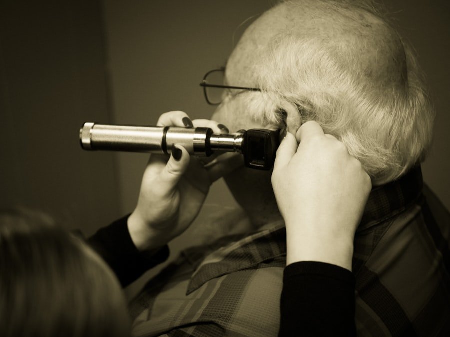
Understanding what happens during your monitoring appointments can help alleviate anxiety and prepare you for what’s to come.
The MRI Appointment: Preparation and Procedure
Your MRI appointments are a routine part of your acoustic neuroma management.
Pre-Appointment Instructions: What to Do Beforehand
You’ll likely receive instructions regarding any necessary dietary restrictions (especially if you’re having contrast), informing the technician of any metal implants or devices you have (as MRI uses magnets), and wearing comfortable clothing without metal components.
Informing About Medical Devices: Safety First
It is crucial to inform the MRI staff about any pacemakers, cochlear implants, aneurysm clips, or other metal prosthetics you may have, as these can interfere with the MRI or pose a safety risk.
Pregnancy and MRI: Important Considerations
If you are pregnant or suspect you might be, you must inform the radiology department before your scan, as MRI is generally considered safe in pregnancy, but precautions are taken.
During the MRI Scan: What Happens in the Machine
The MRI machine is a large, tube-like device. You will lie on a movable table that slides into the scanner. The technician will communicate with you through an intercom system. The scanner produces loud knocking and buzzing noises, which are normal. You may be given earplugs or headphones for comfort. You will be asked to remain as still as possible during the scan to ensure image clarity.
Staying Still: The Key to Clear Images
Movement can blur the images, making them difficult to interpret. Following the technician’s instructions to remain still is paramount for accurate results.
The Importance of Open Communication: Your Comfort and Safety
If you experience claustrophobia or discomfort during the scan, communicating this to the technician is important. There are often open MRI machines available for those with severe claustrophobia, though image quality can sometimes be slightly different.
The Audiology Appointment: Assessing Your Hearing
These appointments focus on evaluating your auditory and sometimes vestibular function.
Hearing Tests by an Audiologist: The Detailed Evaluation
An audiologist will conduct a series of tests, including pure-tone audiometry, speech audiometry, and tympanometry. These tests assess your ability to hear different frequencies and volumes, understand speech, and evaluate the function of your middle ear.
Understanding the Results: What an Audiologist Explains
The audiologist will explain your results, highlighting any changes from previous tests and discussing what these changes might mean in the context of your acoustic neuroma.
Discussing Symptom Impact: Connecting Hearing to Your Life
You’ll have the opportunity to discuss how your hearing or any auditory symptoms are impacting your daily life.
Follow-up with Your Specialist: Integrating the Findings
After your scans and tests, you’ll have a follow-up appointment with your neuro-oncologist or neurosurgeon.
Reviewing Imaging and Test Results: The Comprehensive Picture
Your doctor will meticulously review all your recent MRI scans, audiology reports, and any other relevant test results. They will compare these findings to your previous results to identify any changes or patterns.
Explaining the Findings in Plain Language: Empowering Understanding
They will explain the results to you in clear and understandable terms, outlining whether the tumor has grown, remained stable, or if there are any concerning changes.
Discussing Treatment and Management Options: Tailoring Your Plan
Based on the review of your results and your overall health and preferences, your doctor will discuss the best course of action. This might involve continuing with your current monitoring schedule, adjusting the frequency of your check-ups, or considering treatment options if the tumor has grown significantly or is causing problematic symptoms.
Q&A Session: Your Opportunity to Ask Anything
This is your crucial time to ask any questions you may have. Don’t hesitate to voice any concerns, seek clarification on medical jargon, or discuss any anxieties you might be experiencing.
Never Hesitate to Ask: Your Health is the Priority
No question is too small or insignificant. Your understanding and comfort are paramount for effective management of your acoustic neuroma.
Understanding Next Steps: Clarity on Your Future Care
Ensure you leave the appointment with a clear understanding of your next steps, including the date of your next scheduled check-up and what symptoms you should report immediately.
Factors Influencing Treatment Decisions: When Monitoring Leads to Action
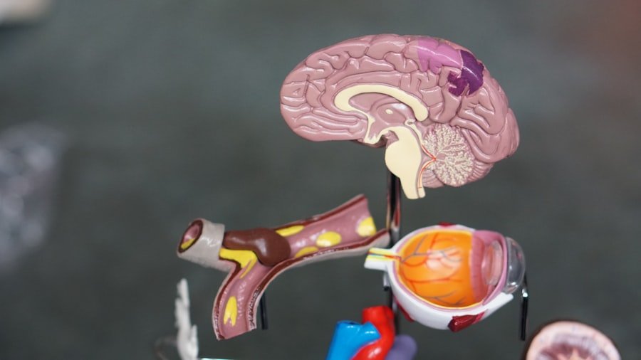
While monitoring is often about observation, it can also be the catalyst for treatment decisions if changes are detected.
Tumor Growth Thresholds: Defining Significant Change
Medical professionals typically have established thresholds for what constitutes “significant” tumor growth that warrants further discussion and potential treatment.
Small Increments of Growth: Usually Managed Conservatively
An acoustic neuroma growing by just a millimeter or two over several years is often considered stable and might not necessitate immediate intervention, especially if it’s not causing symptoms.
Larger or Accelerated Growth: Prompting Treatment Discussions
However, if the tumor shows a more substantial increase in size over a shorter period, or if its growth is causing or likely to cause significant symptoms, treatment options will be actively discussed.
Symptom Development and Severity: The Patient Experience
The emergence or worsening of symptoms is a major driver for considering treatment.
New or Worsening Hearing Loss: A Common Concern
If you experience a significant and rapid deterioration in your hearing, or if your tinnitus becomes unbearable, these symptoms often prompt a re-evaluation of your management plan.
Impact on Daily Functioning: Quality of Life Considerations
The impact of your symptoms on your daily functioning, your work, and your overall quality of life will be a significant factor in treatment decisions.
Balance Issues and Facial Nerve Involvement: Urgent Concerns
Dizziness, vertigo, and any signs of facial nerve weakness (such as facial numbness or paralysis) are often treated more urgently due to their potential to significantly impact quality of life and, in rare cases, indicate more aggressive growth.
Patient Age, Health, and Preferences: A Multifaceted Decision
As discussed earlier, your individual circumstances play a crucial role in deciding whether and when to pursue treatment.
Treatment Risks vs. Benefits: A Careful Calculation
Your doctors will carefully weigh the potential risks and benefits of any proposed treatment against the risks of continued monitoring and potential tumor growth.
Lifestyle and Goals: Aligning Care with Your Life
Your personal goals, lifestyle, and tolerance for potential side effects will be integral to this shared decision-making process.
The Long Haul: Living with an Acoustic Neuroma and Ongoing Surveillance
| Time Period | Frequency of Check-up |
|---|---|
| First 5 years after treatment | Every 6-12 months |
| 5-10 years after treatment | Every 1-2 years |
| After 10 years | Every 2-5 years |
Living with an acoustic neuroma is a marathon, not a sprint. Understanding the long-term implications of monitoring is key to navigating this journey with confidence.
Long-Term Stability and Reduced Monitoring Frequency: A Positive Outcome
For many individuals, their acoustic neuroma will remain stable for years, even decades. In such cases, the frequency of your MRIs and audiology tests will likely decrease over time.
Gradual Lengthening of Intervals: Trusting the Stability
Once your tumor has demonstrated a consistent pattern of stability for several years, your doctor may extend the monitoring intervals from annually to every 18 months or even two years.
Expert Reassurance: Building Confidence in Your Management
This transition signifies a positive outcome – your tumor is not actively growing – and provides reassurance about your ongoing health.
Lifelong Monitoring: A Prudent Approach for Some
While monitoring frequency may decrease, in many cases, some form of lifelong monitoring is recommended, especially for younger individuals or those where the tumor was initially larger.
Continuous Vigilance: Ensuring Early Detection of Any Changes
The rationale behind lifelong monitoring is to ensure that any potential, albeit rare, late-stage growth or changes are detected promptly, allowing for timely intervention if necessary.
The Evolution of MRI Technology: Enhanced Imaging Over Time
As MRI technology continues to advance, scans become more detailed and precise, offering even greater confidence in monitoring even over extended periods.
Transitioning to Symptom Management: When Monitoring Becomes Less Intensive
In certain situations, particularly with very slow-growing tumors in older individuals, the focus might gradually shift from intensive monitoring to managing any existing symptoms.
Prioritizing Quality of Life: Focusing on Well-being
If the tumor is stable, small, and not causing significant symptoms, and if the burdens of frequent monitoring are perceived to outweigh the potential benefits, your doctor might recommend less frequent imaging and a greater emphasis on managing any auditory or vestibular issues.
Balancing Risks and Benefits Over Time: A Dynamic Decision
This decision is always made on a case-by-case basis, carefully considering the individual’s overall health, lifespan, and preferences.
Empowering Yourself Through Knowledge: Your Role in Your Care
As your LCA, my ultimate goal is to empower you with knowledge and understanding. Being informed about your acoustic neuroma and its monitoring process is a vital part of taking an active role in your healthcare.
Open Communication with Your Medical Team: The Cornerstone of Effective Care
Maintain open and honest communication with your neuro-oncologist or neurosurgeon. Don’t hesitate to ask questions, express your concerns, and voice your preferences.
Understanding the “Why”: Demystifying Your Treatment Plan
Understanding why certain checks are recommended will help you feel more confident and compliant with your monitoring schedule.
Staying Informed and Proactive: Be an Advocate for Your Health
Educate yourself about acoustic neuromas and the monitoring process. This knowledge will empower you to be a proactive advocate for your own health and well-being. Remember, you are an integral part of your healthcare team.
FAQs
1. What is acoustic neuroma?
Acoustic neuroma is a non-cancerous tumor that develops on the main nerve leading from the inner ear to the brain.
2. How often should acoustic neuroma be checked?
The frequency of check-ups for acoustic neuroma depends on the size and growth rate of the tumor, as well as the individual’s symptoms and overall health. Generally, regular monitoring through MRI scans is recommended.
3. What are the symptoms of acoustic neuroma?
Symptoms of acoustic neuroma may include hearing loss, ringing in the ear, dizziness, and balance problems. In some cases, the tumor may grow large enough to press against the brain, causing more severe symptoms.
4. What are the treatment options for acoustic neuroma?
Treatment options for acoustic neuroma include observation, radiation therapy, and surgical removal of the tumor. The choice of treatment depends on the size and growth rate of the tumor, as well as the individual’s overall health and preferences.
5. What are the potential complications of acoustic neuroma?
Potential complications of acoustic neuroma include hearing loss, facial weakness or numbness, balance problems, and in rare cases, pressure on the brain leading to more severe neurological symptoms. Regular monitoring and appropriate treatment can help manage these complications.