You’re facing a diagnosis of acoustic neuroma, and the word “surgery” can bring a cascade of questions. As your Listicle Content Architect (LCA), I’m here to demystify the options, breaking down the various surgical approaches available. Understanding these distinct pathways is crucial for making informed decisions alongside your medical team. This isn’t about choosing the “best” surgery in a vacuum, but rather identifying the right surgery for your specific situation. We’ll delve into the primary techniques, their nuances, and what you can expect.
Before we explore the ‘how,’ let’s understand the ‘why.’ Acoustic neuroma surgery, at its core, aims to achieve one or more critical objectives. It’s not a one-size-fits-all solution, and the primary goals shape the surgical approach chosen.
a. Tumor Removal: The Primary Objective
The most common and often primary goal of acoustic neuroma surgery is the complete or partial removal of the tumor. This directly addresses the mass effect the tumor exerts on surrounding nerves and structures, particularly the facial nerve and the cochlear nerve responsible for hearing.
i. Complete Resection: Aiming for Zero Traces
In many cases, the ideal outcome is a complete resection, meaning the surgeon removes the entire tumor. This is often achievable for smaller, well-defined tumors that haven’t significantly invaded critical structures. Complete removal offers the best chance of preventing regrowth and alleviating symptoms caused by tumor pressure. However, the feasibility of complete resection is heavily dependent on the tumor’s size, location, and its relationship with delicate nerves.
ii. Subtotal Resection: When Complete Isn’t Possible
There are instances where a complete tumor removal is not surgically feasible or would carry an unacceptably high risk of damage to vital functions. In such cases, surgeons may opt for a subtotal resection, removing as much of the tumor as safely possible. This can still provide relief from symptoms by reducing the pressure on surrounding nerves. The remaining tumor tissue is often monitored closely with imaging for any signs of growth, and further treatment options like radiation therapy might be considered.
b. Preservation of Nerve Function: A Delicate Balancing Act
While tumor removal is paramount, the preservation of nearby nerve function is an equally, if not more, important consideration for most patients. The acoustic neuroma arises from the Schwann cells of the vestibulocochlear nerve (cranial nerve VIII), which has two main branches: the cochlear nerve (hearing) and the vestibular nerve (balance). In close proximity are other critical nerves, most notably the facial nerve (cranial nerve VII), which controls facial expressions.
i. Hearing Preservation: A Hope for Many
The ability to preserve hearing is a significant factor for many patients, especially those with smaller tumors. Surgical techniques are continually evolving to maximize the chances of saving hearing, though this is not always possible, particularly with larger tumors or when the hearing is already compromised. The decision to prioritize hearing preservation will be influenced by the current state of your hearing, the size and location of the tumor, and the surgeon’s experience.
ii. Facial Nerve Function: A Non-Negotiable Priority
Preserving facial nerve function is almost always a top priority for surgeons, as paralysis or weakness of the face can have a profound impact on a person’s quality of life and immediate appearance. While some degree of risk exists with any surgery in this area, surgeons employ meticulous techniques to identify and meticulously dissect around the facial nerve, minimizing the chances of injury.
iii. Balance Function: Managing the Aftermath
The vestibular nerve plays a crucial role in balance. While its removal is often a consequence of tumor removal, especially for larger tumors, surgeons aim to preserve as much of its function as possible. Even with optimal preservation, some patients may experience temporary or permanent balance issues post-surgery, which can often be managed with rehabilitation.
c. Symptom Relief: Addressing the Distressing Effects
Beyond the direct removal of the tumor, surgery aims to alleviate the often distressing symptoms caused by the tumor’s growth. These symptoms can include hearing loss, tinnitus (ringing in the ear), dizziness or vertigo, and sometimes facial numbness or weakness. Successful surgery can lead to a significant reduction or elimination of these symptoms, improving a patient’s overall well-being.
i. Alleviating Hearing Loss and Tinnitus
The pressure from an acoustic neuroma can compress the cochlear nerve, leading to progressive hearing loss and often a persistent tinnitus. Removing the tumor can relieve this pressure, potentially halting further hearing degradation and, in some cases, even leading to a slight improvement in hearing or a reduction in tinnitus.
ii. Controlling Dizziness and Vertigo
The vestibular nerve, responsible for balance, can also be affected by the tumor. This can manifest as episodic or constant dizziness and vertigo. Tumor removal can alleviate these symptoms by decompressing the vestibular system, providing much-needed stability.
iii. Managing Facial Nerve Symptoms
While less common than hearing or balance issues, larger tumors can sometimes press on the facial nerve, causing subtle weakness or numbness. Surgery can relieve this pressure, preventing further progression of these symptoms.
2. Craniotomy-Based Approaches: The Traditional Routes
These surgical techniques involve opening a portion of the skull to access the tumor. They are often employed for larger tumors or those that have a significant portion extending into the skull base. While invasive, they offer excellent visualization and the ability to remove large tumors.
a. Retrosigmoid (or Retrosigmoid-Cerebellopontine Angle) Approach: A Direct Path
This is a frequently used method, especially for larger tumors. It involves making an incision behind the ear, exposing the mastoid bone, and then drilling away a small portion of the bone to access the cerebellopontine angle (CPA), where the tumor is located.
i. Surgical Incision and Bone Removal: The Entry Point
The incision is typically made behind the mastoid bone, the prominent bone behind your ear. The surgeon then carefully drills away a small section of this bone to gain access to the dura mater, the tough outer covering of the brain.
ii. Tumor Dissection and Removal: Navigating the Nerves
Once the CPA is accessed, the surgeon meticulously identifies and dissects the tumor away from the sensitive cranial nerves, particularly the facial and cochlear nerves. The goal is to remove the entire tumor while preserving these vital structures. This approach offers a good view of the entire cerebellopontine angle, making it suitable for larger tumors.
iii. Advantages and Disadvantages: Weighing the Options
- Advantages: Excellent visualization of the tumor and surrounding structures, allowing for potentially complete resection of large tumors. Relatively good rates of hearing preservation for appropriately selected patients.
- Disadvantages: Requires an incision behind the ear and drilling of bone. There is a risk of CSF leak, meningitis, or damage to nearby blood vessels. Post-operative recovery might involve more discomfort due to the incision site.
b. Translabyrinthine Approach: Prioritizing Facial Nerve Preservation
This approach is favored when hearing preservation is not a primary concern, but preserving facial nerve function is paramount. It involves removing a portion of the bone within the inner ear (labyrinth) to access the tumor.
i. Surgical Incision and Inner Ear Access: A Different Route
The incision is similar to the retrosigmoid approach, behind the ear. However, instead of drilling through the mastoid bone to reach the CPA directly, the surgeon enters the inner ear structures (cochlea and semicircular canals). These are removed to gain access to the tumor.
ii. Tumor Dissection and Facial Nerve Focus: A Specialized Technique
By accessing the tumor through the inner ear, surgeons can often achieve a very direct line of sight to the facial nerve, allowing for meticulous dissection and a high likelihood of preserving its function. Hearing preservation is sacrificed with this approach due to the removal of the inner ear structures.
iii. Advantages and Disadvantages: When Hearing Isn’t the Priority
- Advantages: Excellent for preserving facial nerve function. Effective for removing larger tumors. Can avoid retraction of the cerebellum, which is sometimes necessary in the retrosigmoid approach.
- Disadvantages: Results in complete hearing loss in the operated ear. There is a risk of CSF leak and meningitis. Can lead to post-operative balance issues.
c. Suboccipital Approach: For Tumors Extending Downwards
This approach involves an incision at the back of the head, below the occipital bone. It’s typically used for tumors that extend downwards or are located more medially in the posterior fossa.
i. Surgical Incision and Posterior Fossa Exposure: Reaching Deep
The incision is made at the back of the head, and a portion of the occipital bone is removed to expose the posterior fossa of the skull. This allows direct access to tumors located in this region.
ii. Tumor Dissection with a Posterior View: A Different Perspective
The surgeon works from a posterior perspective, dissecting the tumor away from the brainstem and cranial nerves. This approach can be advantageous for tumors that have a significant extension towards the brainstem.
iii. Advantages and Disadvantages: A Specific Target
- Advantages: Good for tumors with medial or downward extension. May offer a different angle of approach for certain tumor shapes and sizes.
- Disadvantages: Can involve retraction of the cerebellum. Potential risks include CSF leak and damage to brainstem structures. Hearing preservation is often challenging with this approach.
3. Endoscopic Ear Surgery: Minimally Invasive Precision
This is a more modern approach that utilizes an endoscope – a small, flexible tube with a camera – to visualize and remove the tumor through the ear canal. It is generally best suited for smaller tumors.
a. The Endoscopic Tool: A Tiny Camera, Big Impact
The endoscope allows surgeons to see inside the body with magnified clarity without the need for a large incision. This minimally invasive nature is the hallmark of this technique.
i. Endoscope Insertion and Visualization: Peering Inside
A small endoscope is inserted through the ear canal. The camera transmits high-definition images to a monitor, providing the surgeon with a detailed view of the tumor and surrounding structures.
ii. Specialized Instruments: Working Through Small Openings
Tiny, specialized surgical instruments are passed through the ear canal alongside the endoscope. These instruments are used to carefully dissect and remove the tumor.
b. Advantages of Endoscopic Approaches: Less Invasion, Faster Recovery
The primary benefits of endoscopic surgery stem from its minimally invasive nature.
i. Reduced Scarring and Pain: A Gentler Recovery
Because the access is through the ear canal, there are typically no visible external scars. This translates to less post-operative pain and a generally more comfortable recovery period.
ii. Shorter Hospital Stays and Quicker Return to Activities: Back to Life Faster
Patients often experience shorter hospital stays and can return to their normal activities more quickly compared to open craniotomy procedures. This is a significant advantage for many individuals.
iii. Potential for Improved Facial Nerve Preservation: Delicate Precision with Visualization
The enhanced visualization provided by the endoscope can allow for more precise dissection around the facial nerve, potentially leading to better preservation of its function, especially for smaller tumors where the nerve might be more easily identified and protected.
c. Limitations and Considerations: When It’s Not the Right Fit
While promising, endoscopic surgery is not always the optimal choice for every acoustic neuroma.
i. Tumor Size and Location Restrictions: Not for Every Tumor
This technique is generally best suited for smaller, well-defined tumors. Larger tumors that have grown significantly or have extensive involvement may not be amenable to removal solely via an endoscopic approach.
ii. Potential for Hearing Loss: A Trade-off to Consider
While the goal is always to preserve function, hearing preservation can be more challenging with the endoscopic approach for certain tumor locations within the cerebellopontine angle. The surgeon will assess this risk carefully.
iii. Surgeon Expertise: A Specialized Skill Set
Performing endoscopic ear surgery requires specialized training and expertise. Not all surgeons are equally experienced in this technique, so finding a surgeon proficient in this approach is crucial.
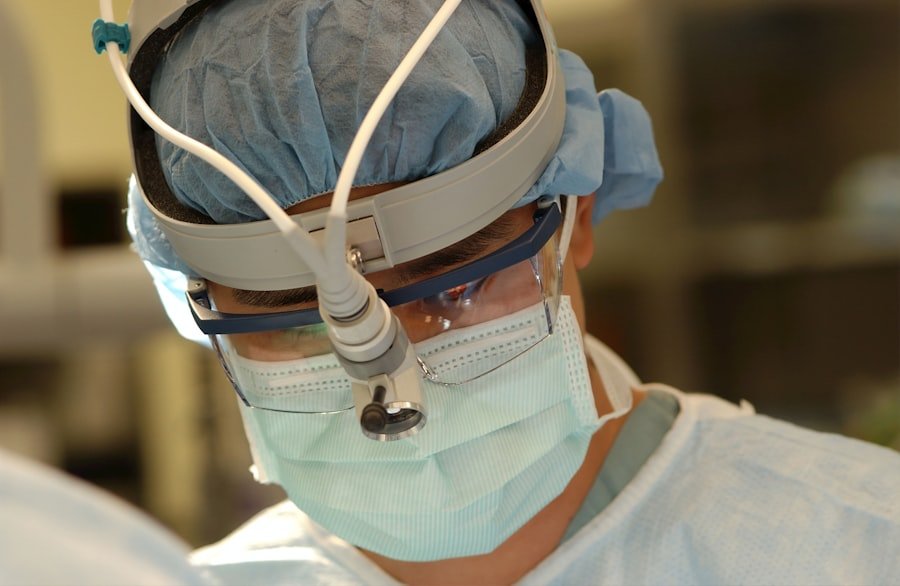
4. Vestibular Schwannoma Micro-Surgery: The Gold Standard of Open Procedures

This category encompasses the open surgical techniques like the retrosigmoid and translabyrinthine approaches, often referred to collectively as microsurgery because of the use of microscopes and delicate instruments. The term “micro-surgery” emphasizes the meticulous, precise dissection performed under magnification.
a. The Surgeon’s Toolkit: Magnification is Key
The cornerstone of these open procedures is the surgical microscope. This allows the surgeon to visualize structures at a magnified level, essential for navigating the delicate neural pathways.
i. Surgical Microscope: The Window to Precision
This high-powered microscope provides a magnified, stereoscopic view of the surgical field. It’s indispensable for identifying and meticulously dissecting around the cranial nerves, blood vessels, and the tumor itself.
ii. Specialized Instruments: Tools for Delicate Work
Along with the microscope, surgeons use a range of specialized, fine instruments – micro-dissectors, micro-forceps, micro-suction devices – designed for precise manipulation and removal of tissue in a confined surgical space.
b. The Operative Process: A Step-by-Step Breakdown
While each approach has its specific steps, the overarching philosophy of microsurgery remains consistent: careful identification, meticulous dissection, and controlled removal.
i. Anesthesia and Incision: Setting the Stage
The patient is placed under general anesthesia. An incision is made based on the chosen approach (e.g., behind the ear).
ii. Bone Removal and Dura Opening: Creating Access
A portion of bone is carefully removed to expose the cerebellopontine angle or inner ear structures. The dura mater is then opened to access the brain.
iii. Tumor Identification and Dissection: The Delicate Dance
Under the microscope, the tumor is meticulously identified. The surgeon then painstakingly dissects the tumor away from the surrounding nerves and blood vessels, often using gentle aspiration and micro-dissection techniques.
iv. Tumor Removal and Closure: Completing the Procedure
Once the tumor is removed, the dura is closed, the bone is replaced (if applicable), and the incision is sutured.
c. Outcomes and Recovery: What to Expect Post-Surgery
Recovery from microsurgery is a process that involves healing, symptom management, and rehabilitation.
i. Immediate Post-Operative Period: The Initial Recovery
Patients are closely monitored in the intensive care unit initially. Pain management, monitoring for neurological changes, and preventing complications like CSF leaks are paramount.
ii. Short-Term Recovery: Weeks to Months
Hospital stays vary but typically range from a few days to a week. Patients will experience fatigue and may have some balance issues or dizziness. Gradual return to daily activities is encouraged.
iii. Long-Term Recovery and Rehabilitation: Regaining Function
Full recovery can take several months to over a year. Physical and occupational therapy are often crucial for regaining balance, coordination, and managing any residual hearing loss or facial nerve weakness. Regular follow-up appointments with imaging scans are essential to monitor for any tumor recurrence.
5. Radiosurgery Techniques: A Non-Invasive Alternative to Surgery
| Surgery Type | Description | Success Rate |
|---|---|---|
| Microsurgery | A surgical procedure using a microscope to remove the tumor | 80-90% |
| Radiation Therapy | Using high-energy radiation to shrink or destroy the tumor | 70-90% |
| Stereotactic Radiosurgery | A non-invasive radiation therapy delivered in a single high dose | 80-90% |
While not strictly “surgery” in the traditional sense of tissue removal with scalpels, stereotactic radiosurgery is a crucial part of the acoustic neuroma treatment landscape. It uses precisely targeted radiation to control tumor growth without making any incisions.
a. Stereotactic Radiosurgery (SRS): Precision Radiation
SRS delivers highly focused beams of radiation to the tumor. The “stereotactic” aspect refers to the use of a system that precisely maps the tumor’s location in three dimensions, ensuring that the radiation is directed only to the tumor and spares surrounding healthy tissue.
i. Gamma Knife Radiosurgery: The Pioneering System
The Gamma Knife is a well-established SRS system that uses multiple cobalt-60 radiation sources arranged in a hemisphere. These beams converge precisely on the tumor, delivering a high dose of radiation while minimizing exposure to healthy brain tissue.
ii. Linear Accelerator (LINAC) Based Radiosurgery: Another Powerful Tool
LINAC-based radiosurgery uses a machine that generates radiation beams. Similar to the Gamma Knife, it employs sophisticated imaging and planning systems to deliver precise radiation doses to the tumor. This approach can be more flexible in terms of beam shaping.
b. CyberKnife Radiosurgery: Robotic Precision
The CyberKnife is another advanced radiosurgery system that utilizes a robotic arm to deliver radiation. Its unique feature is its ability to track the tumor’s movement in real-time (e.g., due to breathing) and adjust the radiation beams accordingly.
i. Real-Time Tumor Tracking: Adapting to Movement
This dynamic tracking capability is a significant advantage, especially for tumors that might move slightly during the treatment session. It ensures that the radiation remains precisely focused on the target.
ii. Advanced Treatment Planning: Tailoring the Dose
CyberKnife offers sophisticated treatment planning software that allows for highly customized radiation delivery, optimizing the dose to the tumor while sparing surrounding critical structures.
c. Advantages of Radiosurgery: The Non-Incisional Benefits
The appeal of radiosurgery lies in its non-invasive nature, offering a compelling alternative for specific patients.
i. No Incisions, No Anesthesia: A Less Traumatic Experience
The absence of incisions and anesthesia makes radiosurgery a significantly less traumatic experience for patients compared to open surgery. This can be particularly appealing for those who are not good surgical candidates or are hesitant about invasive procedures.
ii. Minimal Recovery Time and Rapid Return to Normal Activities: Back to Life Immediately
Since there is no surgery, the recovery time is virtually non-existent. Patients can usually resume their normal routines almost immediately after the treatment session.
iii. High Success Rates in Tumor Control: Slowing Down Growth
Radiosurgery has a high success rate in controlling the growth of acoustic neuromas. While it doesn’t typically remove the tumor, it effectively stops it from growing larger, thereby alleviating symptoms caused by pressure.
iv. Preservation of Hearing and Facial Nerve Function: A Key Advantage
A major benefit of radiosurgery is its excellent track record in preserving hearing and facial nerve function, especially for smaller tumors. The targeted nature of the radiation minimizes the risk of damage to these delicate nerves.
d. Limitations and Considerations: When Radiosurgery Might Not Be the Best Option
Despite its advantages, radiosurgery is not a universal solution for all acoustic neuromas.
i. Tumor Size and Location: Not for Every Tumor
Radiosurgery is generally most effective for smaller tumors that are well-defined and not impinging significantly on critical structures. Larger tumors may require surgery for effective removal or to alleviate immediate pressure symptoms.
ii. Tumor Growth and Aggressiveness: Different Approaches for Different Tumors
While radiosurgery is excellent for control, it may not be the best option for rapidly growing or highly aggressive tumors. In such cases, surgical removal might be preferred to address the tumor burden more directly.
iii. Long-Term Monitoring is Still Necessary: Ongoing Vigilance
Although radiosurgery effectively halts tumor growth, regular MRI scans are still necessary to monitor the tumor size and ensure there is no recurrence or residual growth. The effects of radiation can take time to become fully apparent, and long-term follow-up is crucial.
You’ve now traversed the landscape of acoustic neuroma surgery. Each technique offers a distinct pathway, with its own set of advantages, disadvantages, and ideal candidates. Your journey from diagnosis to treatment is a collaborative one, and by arming yourself with this knowledge, you are empowered to engage in meaningful discussions with your neurosurgeon and audiologist, ultimately guiding you to the surgical intervention that best aligns with your health, your tumor characteristics, and your life goals.
FAQs
What is an acoustic neuroma?
An acoustic neuroma is a non-cancerous tumor that develops on the main nerve leading from the inner ear to the brain. It can affect hearing and balance.
What are the types of acoustic neuroma surgery?
There are three main types of acoustic neuroma surgery: microsurgery, radiosurgery, and endoscopic surgery. Each type has its own benefits and risks.
What is microsurgery for acoustic neuroma?
Microsurgery involves removing the tumor through a small incision behind the ear using a microscope and specialized instruments. This allows for precise tumor removal while preserving surrounding nerves.
What is radiosurgery for acoustic neuroma?
Radiosurgery, also known as stereotactic radiosurgery, uses highly focused radiation beams to target and destroy the tumor. This type of surgery is non-invasive and does not require any incisions.
What is endoscopic surgery for acoustic neuroma?
Endoscopic surgery involves using a small camera and specialized instruments to remove the tumor through the nasal cavity. This approach may be suitable for smaller tumors and can result in less scarring and a shorter recovery time.