You’re about to embark on a journey to understand a complex medical procedure: acoustic neuroma surgery. As the Listicle Content Architect, my goal is to break down this intricate process into digestible, informative sections, ensuring you grasp every crucial detail. This isn’t just about listing facts; it’s about guiding you through the labyrinth of what happens, from preparation to recovery, with clarity and comprehensive insight. Get ready to gain a profound understanding of how this surgery addresses the acoustic neuroma.
Before you even enter the operating room, a significant amount of planning and preparation takes place. This phase is critical, as it ensures the surgical team has all the necessary information and that you are in the best possible condition for the procedure. Think of it as building the foundation before constructing the skyscraper – absolutely essential for stability and success.
A. Comprehensive Medical Evaluations and Imaging
Your journey begins with a thorough assessment of your overall health. This isn’t just a cursory check; it’s a deep dive into your medical history, existing conditions, and any medications you’re currently taking.
- History and Physical Exam: The neurosurgeon or otolaryngologist (ENT surgeon) will meticulously review your medical history. They’ll ask detailed questions about your symptoms, their onset, and progression. This includes understanding any hearing loss, tinnitus, dizziness, or facial nerve issues you might be experiencing. A physical examination will further assess your general health and identify any potential risk factors.
- Neurological Assessment: This is a vital part of the evaluation, focusing on your cranial nerves, balance, and coordination. The surgeon will test your hearing, facial sensation and movement, and gait to establish a baseline for comparison after surgery.
- Imaging Studies: This is where we get a clear picture of the acoustic neuroma.
- MRI (Magnetic Resonance Imaging): This is the gold standard for diagnosing acoustic neuromas. A gadolinium-enhanced MRI provides highly detailed images of the tumor, its size, location, and its relationship to surrounding critical structures like the brainstem, cerebellum, and cranial nerves (especially the facial and auditory nerves). You’ll likely have multiple MRI scans to track tumor growth over time if surgery is not immediately recommended, and for precise surgical planning.
- CT (Computed Tomography) Scan: While MRI is preferred for soft tissue detail, a CT scan might be used in certain situations, perhaps to assess bony structures or if an MRI is contraindicated.
B. Pre-Surgical Consultations and Team Assembly
Surgery is a team sport, and acoustic neuroma removal is no exception. You’ll meet with various specialists who will play a role in your care.
- Surgeon Consultation: This is your primary point of contact. The surgeon will explain the different surgical approaches, discuss the risks and benefits specific to your case, and answer all your questions. They will review your imaging and explain what they expect to find during the surgery.
- Anesthesiologist Consultation: The anesthesiologist will discuss the type of anesthesia that will be used (usually general anesthesia) and assess your suitability for it. They will outline the anesthetic plan and address any concerns you have about pain management or sedation.
- Audiologist Evaluation: If hearing loss is a significant symptom, an audiologist will perform comprehensive hearing tests. This helps document your current hearing level, which is crucial for assessing potential hearing preservation or loss after surgery.
- Neuromonitoring Team: For acoustic neuroma surgery, real-time monitoring of nerve function is often employed. You’ll meet the neuromonitoring technician who will explain how they will track the electrical activity of your facial and auditory nerves during the procedure to help the surgeon avoid injury.
C. Pre-Operative Instructions and Preparations
To ensure the surgery proceeds smoothly and safely, you’ll receive specific instructions.
- Medication Review: You’ll be asked to provide a complete list of all medications, including over-the-counter drugs, supplements, and herbal remedies. Certain medications, like blood thinners, may need to be stopped before surgery to reduce the risk of bleeding.
- Fasting Instructions: You will be instructed not to eat or drink for a certain period before surgery, typically starting at midnight the night before. This is standard practice for any surgical procedure under general anesthesia.
- Hygiene and Skin Preparation: You might be asked to shower with a special antiseptic soap to reduce the risk of infection. Any hair in the surgical area may be clipped, but usually not fully shaved at this stage.
- Logistics and Support: Arrange for someone to drive you to and from the hospital and to stay with you for the initial period of recovery. Discuss the hospital admission process and what items you should and should not bring with you.
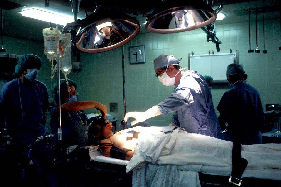
2. The Surgical Arena: Navigating the Procedure
Now, you’re at the hospital, and it’s time for the surgery itself. This is where the medical team utilizes their expertise and advanced technology to access and remove the acoustic neuroma. The choice of surgical approach depends on several factors, including the size and location of the tumor, your hearing status, and the surgeon’s preference.
A. Anesthesia and Patient Positioning
The moment you enter the operating room, your comfort and safety are paramount.
- Induction of Anesthesia: You will be taken into the operating room, and the anesthesiologist will administer the anesthetic. This will cause you to fall into a deep, unconscious sleep, ensuring you feel no pain or discomfort during the procedure.
- Vascular Access: An intravenous (IV) line will be placed, usually in your arm or hand. This line will be used to administer fluids, medications, and anesthesia throughout the surgery.
- Monitoring Equipment: A variety of monitors will be attached to you to continuously track your vital signs, including your heart rate, blood pressure, oxygen saturation, and temperature. This ensures your body is responding well to the anesthesia and the surgical stress.
- Patient Positioning: The way you are positioned on the operating table is crucial for providing the surgeon with optimal access to the tumor while protecting surrounding structures. The specific position varies depending on the surgical approach:
- Supine (Lying on your back): Often used for translabyrinthine and retrosigmoid approaches, sometimes with the head turned and slightly extended.
- Prone (Lying on your stomach): Less common for acoustic neuroma surgery but can be used in certain complex cases.
- Lateral (Lying on your side): Can be employed for some approaches.
The surgical team will ensure you are comfortably and securely positioned to prevent pressure sores or nerve damage.
B. The Surgical Approaches: Pathways to the Tumor
There are several well-established surgical techniques for removing acoustic neuromas, each with its own advantages and disadvantages.
- Retrosigmoid Approach (Posterior Cranial Fossa Approach):
- Incision: A small incision is made behind the ear, extending towards the midline of the back of the head.
- Craniotomy: A small opening in the skull (craniotomy) is made in the posterior cranial fossa, allowing access to the tumor.
- Advantages: This approach offers good visualization of the tumor, brainstem, and cranial nerves. It often allows for the best chance of hearing preservation in tumors where this is a possibility.
- Considerations: It requires careful dissection near the cerebellum and brainstem.
- Translabyrinthine Approach:
- Incision: An incision is made in front of and behind the ear, extending into the ear canal.
- Skull Base Opening: The surgeon removes bone from the mastoid bone (behind the ear) and then enters the inner ear (labyrinth) to access the tumor.
- Advantages: This approach provides excellent access to tumors that extend into the internal auditory canal and cerebellopontine angle. It is often chosen when hearing preservation is unlikely or has already been lost.
- Considerations: This approach inevitably sacrifices hearing in the operated ear.
- Middle Fossa Approach:
- Incision: An incision is made above the ear.
- Skull Base Opening: A small craniotomy is performed in the middle fossa of the skull.
- Advantages: This approach offers direct visualization of the superior aspect of the internal auditory canal and can be good for smaller tumors. It may allow for better preservation of facial nerve function in certain cases.
- Considerations: It can be more challenging for larger tumors or those extending further.
C. Tumor Removal and Nerve Preservation
This is the core of the surgical intervention. The surgeon’s skill and the use of advanced tools are critical here.
- Microsurgical Techniques: The surgery is performed under high magnification using a surgical microscope. This allows the surgeon to see tiny structures with great clarity. Specialized micro-instruments are used for dissection.
- Identification and Dissection: The surgeon meticulously identifies the acoustic neuroma and carefully dissects it away from the surrounding brain tissue, brainstem, and vital cranial nerves. This is a delicate process, as the tumor can be intimately involved with these structures.
- Facial Nerve Monitoring: Throughout the dissection, the facial nerve (which controls facial expressions) is continuously monitored. Electrodes are placed near the nerve, and the neuromonitoring team provides real-time feedback to the surgeon regarding the nerve’s function. If the nerve shows signs of compromise, the surgeon will adjust their technique to minimize the risk of injury.
- Auditory Nerve Monitoring: Similar monitoring can be performed for the auditory nerve, though it is often more challenging and may not always be successful in preserving hearing.
- Tumor Resection: The acoustic neuroma is gradually removed. Depending on the size and location of the tumor, the surgeon may aim for:
- Subtotal Resection: Removing as much of the tumor as possible while preserving critical nerves. This might be chosen if the tumor is very large and tightly adhered to vital structures.
- Total Resection: Removing the entire tumor. This is the goal when possible, especially for smaller tumors.
- Intraoperative Imaging: In some cases, intraoperative MRI or ultrasound may be used to confirm complete tumor removal and ensure no residual tumor remains.
D. Closure and Dressings
Once the tumor removal is complete, the surgical site is meticulously closed.
- Hemostasis: The surgical field is carefully reviewed to ensure all bleeding has stopped. This is crucial to prevent post-operative hematomas (collections of blood).
- Dural Closure: The dura mater, the tough membrane surrounding the brain, is carefully repaired if it was opened.
- Bone Flap Replacement (if applicable): If a bone flap was created during the craniotomy, it is repositioned and secured in place.
- Layered Closure: The overlying tissues – muscles, subcutaneous tissue, and skin – are then closed in layers with sutures or staples.
- Dressings: A sterile dressing will be applied to the surgical site to protect it and absorb any drainage.
3. Post-Operative Recovery: The Healing Process Begins

The moment you wake up from anesthesia marks the beginning of your recovery journey. This phase is characterized by careful monitoring, pain management, and gradual rehabilitation.
A. Immediate Post-Operative Care in the ICU or PACU
You will spend the initial hours after surgery in a specialized unit designed for close observation.
- Recovery from Anesthesia: You’ll be closely monitored as you wake up from the general anesthesia. The anesthesia team will ensure your vital signs are stable.
- Pain Management: Pain medication will be administered to keep you comfortable. This may include intravenous medications initially, transitioning to oral medications as you become more alert.
- Neurological Assessments: Frequent neurological checks will be performed by the nursing staff and physicians. This includes assessing your level of consciousness, eye movements, facial muscle strength, and any sensory changes.
- Monitoring for Complications: The medical team will be vigilant for any signs of complications such as bleeding, infection, or swelling.
- Fluid Management: Intravenous fluids will be administered to maintain hydration and electrolyte balance.
B. Transfer to a Regular Hospital Room and Initial Mobilization
Once you are stable and your condition allows, you’ll be moved to a regular hospital room.
- Mobility: Your care team will encourage you to start moving as soon as it’s safe. This might begin with sitting up in bed, progressing to short walks with assistance. Early mobilization is crucial to prevent blood clots and pneumonia.
- Dietary Progression: You’ll likely start with clear liquids and gradually advance to solid foods as tolerated, depending on your bowel function and overall comfort.
- Wound Care: Staples or sutures will be monitored for any signs of infection or drainage. The dressing will be changed as needed. You’ll be instructed on how to care for the surgical wound at home.
- Medication Regimen: You’ll continue to take prescribed medications for pain, and potentially to prevent blood clots or manage other conditions.
C. Addressing Common Post-Operative Symptoms
It’s normal to experience certain effects following acoustic neuroma surgery. Understanding these can help you cope with them.
- Dizziness and Balance Issues: Dizziness is very common, especially after translabyrinthine surgery, as the inner ear structures have been affected. This can range from mild lightheadedness to significant vertigo. Physiotherapy and specific exercises will be incorporated into your rehabilitation to help your brain adapt.
- Hearing Changes: You may experience further hearing loss in the operated ear, especially with approaches that involve the inner ear. Tinnitus (ringing in the ear) might also be present or worsen.
- Facial Weakness or Numbness: Temporary or, less commonly, permanent weakness or numbness in the face can occur due to manipulation or stretching of the facial nerve during surgery. This often improves over time with rehabilitation.
- Headaches: Post-operative headaches are common and are usually managed with medication.
- Fatigue: It’s normal to feel very tired for several weeks or even months after surgery as your body heals.
D. Rehabilitation and Therapy Services
A key component of your recovery involves regaining function and adapting to any changes.
- Physical Therapy: This is crucial for addressing balance and coordination issues. Therapists will guide you through exercises to improve your gait, stability, and vestibular function.
- Occupational Therapy: If fine motor skills or daily living activities are affected, an occupational therapist can provide strategies and exercises to help you regain independence.
- Speech and Swallowing Therapy: In some cases, you might experience difficulty with swallowing or speaking if cranial nerves affecting these functions have been impacted. A speech-language pathologist can assist with this.
- Audiology and Hearing Rehabilitation: If hearing loss is significant, an audiologist can assess your hearing and discuss options such as hearing aids, cochlear implants, or other assistive listening devices.
4. Potential Complications and Their Management
While acoustic neuroma surgery is generally safe and effective, like any major surgical procedure, there are potential risks and complications. Being aware of these can empower you to communicate any concerns to your medical team.
A. Neurological Deficits
Damage to critical nerves is a primary concern.
- Facial Nerve Palsy: As mentioned, this can range from temporary weakness to permanent paralysis of the facial muscles. Management involves vigilant monitoring during surgery, early recognition of any deficit, and rehabilitation (physical therapy, sometimes medications like corticosteroids).
- Hearing Loss: Even with attempts to preserve hearing, some degree of loss is common, particularly with translabyrinthine approaches. In rare cases, hearing can be worsened or completely lost.
- Balance Impairment (Vertigo): This is a common, often temporary, side effect. Severe or persistent vertigo may require specialized vestibular rehabilitation therapy.
- Cranial Nerve V (Trigeminal Nerve) Issues: This nerve provides sensation to the face. Injury can lead to facial numbness, tingling, or pain.
- Other Cranial Nerve Issues: Less commonly, other cranial nerves might be affected, leading to issues with swallowing, voice, or shoulder movement.
B. Cerebrospinal Fluid (CSF) Leak
The brain is surrounded by cerebrospinal fluid. If the dura mater (the membrane protecting the brain) is not perfectly sealed, CSF can leak out.
- Symptoms: A CSF leak typically presents as a clear, watery discharge from the nose or ear, often accompanied by headaches that worsen when upright.
- Management: Small leaks may resolve on their own with bed rest. Larger or persistent leaks may require further intervention, such as a lumbar drain to relieve pressure or surgical repair of the dural defect.
C. Infection
Any surgical site carries a risk of infection.
- Symptoms: Signs of infection can include fever, increased redness, swelling, pain at the surgical site, or discharge.
- Management: Infections are typically treated with antibiotics. In some cases, further surgery may be needed to clean out the infected area.
D. Bleeding and Hematoma
| Stage | Procedure |
|---|---|
| Pre-surgery | Medical evaluation, imaging tests, and discussion with the surgical team |
| Anesthesia | General anesthesia is administered to the patient |
| Incision | A small incision is made behind the ear to access the skull base |
| Tumor removal | The acoustic neuroma is carefully removed from the nerves in the inner ear |
| Nerve preservation | Efforts are made to preserve facial nerve function and hearing |
| Closure | The incision is closed with sutures or staples |
| Recovery | Patient is monitored in the recovery room before being transferred to a hospital room |
Bleeding can occur during or after surgery. A significant collection of blood is called a hematoma.
- Risk: This is why meticulous hemostasis during surgery is so important.
- Management: Small hematomas may be reabsorbed by the body. Larger hematomas can cause pressure on the brain and may require surgical drainage.
E. Anesthesia-Related Complications
While rare, complications related to anesthesia can occur.
- Allergic Reactions: The anesthesiologist will screen for allergies before administering any medications.
- Respiratory Issues: Problems with breathing during or after anesthesia.
- Nausea and Vomiting: Common side effects that are usually managed with medication.
F. Tumor Recurrence or Residual Tumor
In some cases, a small portion of the tumor might be left behind (residual tumor), or the tumor may grow back over time (recurrence).
- Monitoring: Regular follow-up imaging with MRI is crucial to detect any signs of residual or recurrent tumor.
- Management: If detected, further treatment options, such as observation, repeat surgery, or radiation therapy, will be discussed.
5. Long-Term Outlook and Follow-Up Care
Your journey doesn’t end when you leave the hospital. Acoustic neuroma surgery requires ongoing monitoring and adaptation to ensure the best possible long-term outcome.
A. Regular Medical Follow-Up and Imaging
Consistent check-ups are vital for monitoring your recovery and detecting any potential issues early.
- Post-Operative Appointments: You will have scheduled appointments with your surgeon at regular intervals after discharge. These appointments allow the surgeon to assess your recovery, monitor for any complications, and evaluate your neurological status.
- MRI Scans: Magnetic Resonance Imaging (MRI) scans are essential for monitoring the surgical site. Initially, these scans will be done more frequently (e.g., every 6 months to a year), and then the interval may be extended to every 1-2 years or as deemed necessary by your physician. These scans help to:
- Confirm complete tumor removal.
- Detect any signs of residual tumor.
- Identify any recurrence of the tumor.
- Assess for any post-operative changes in the surrounding brain tissue.
- Audiology Appointments: If hearing preservation was a goal or if hearing loss is a significant concern, regular audiology appointments are crucial. These will involve hearing tests to track any changes in your hearing ability and to discuss management strategies.
B. Lifestyle Adjustments and Support Systems
Adapting to life after acoustic neuroma surgery often involves making some lifestyle adjustments and leaning on your support network.
- Patience and Self-Care: Recovery from acoustic neuroma surgery can be a lengthy process. It’s important to be patient with yourself and prioritize rest, adequate nutrition, and hydration. Avoid overexertion and listen to your body.
- Managing Residual Symptoms: If you experience persistent dizziness, balance issues, or facial weakness, continue with your prescribed rehabilitation exercises. Don’t hesitate to communicate with your healthcare team about any ongoing challenges. They can offer further strategies or referrals.
- Emotional and Psychological Well-being: Undergoing brain surgery can be a stressful and emotional experience. It’s normal to experience anxiety, frustration, or even depression.
- Support Groups: Connecting with others who have been through similar experiences can be incredibly beneficial. Many hospitals and patient advocacy groups offer support networks, both in-person and online.
- Counseling or Therapy: If you are struggling with your emotional well-being, consider seeking professional help from a therapist or counselor. They can provide coping mechanisms and support.
- Family and Friends: Lean on your loved ones for emotional support and practical assistance. They can be invaluable in helping you navigate the recovery period.
C. Return to Work and Daily Activities
The timeline for returning to work and resuming normal activities varies significantly from person to person.
- Gradual Reintegration: Most individuals can gradually return to work and their usual activities as their strength and energy levels improve.
- Doctor’s Clearance: Your surgeon will provide guidance on when it is safe to resume specific activities, such as driving or strenuous exercise.
- Accommodation: If you experience ongoing challenges, discuss potential accommodations with your employer to ensure a smooth transition back into the workforce. This might include modified work schedules or a less demanding role initially.
D. Understanding the Long-Term Prognosis
The outlook for individuals who have undergone acoustic neuroma surgery is generally very positive, especially for smaller tumors.
- Benign Nature: Acoustic neuromas are almost always benign (non-cancerous) tumors.
- Successful Removal: When completely removed, the risk of recurrence is low.
- Quality of Life: With effective management of any residual symptoms and ongoing medical care, most individuals can maintain a good quality of life after surgery. The focus shifts from fighting the tumor to managing its after-effects and embracing a healthy lifestyle.
- Early Detection and Intervention: The continued advancement in diagnostic imaging and surgical techniques means that acoustic neuromas are often detected and treated earlier, leading to even better outcomes.
As you navigate the complexities of acoustic neuroma surgery, remember that you are not alone. You are at the center of a dedicated medical team, and with thorough preparation, skilled surgical intervention, and dedicated post-operative care, the path to recovery and a fulfilling life is well within reach. This detailed exploration aims to equip you with the knowledge and understanding to feel confident and prepared every step of the way.
FAQs
What is an acoustic neuroma?
An acoustic neuroma is a non-cancerous tumor that develops on the main nerve leading from the inner ear to the brain. It can cause hearing loss, ringing in the ear, and unsteadiness.
When is surgery recommended for acoustic neuroma?
Surgery is typically recommended for acoustic neuroma if the tumor is large, growing rapidly, or causing significant symptoms such as hearing loss, balance problems, or facial numbness.
What happens during acoustic neuroma surgery?
During acoustic neuroma surgery, the surgeon will make an incision behind the ear, remove a small piece of the skull, and carefully separate the tumor from the surrounding nerves and blood vessels. The goal is to remove as much of the tumor as possible without damaging the surrounding structures.
What are the risks of acoustic neuroma surgery?
Risks of acoustic neuroma surgery include hearing loss, facial weakness, balance problems, and damage to the nerves that control facial movement and sensation. There is also a risk of infection, bleeding, and complications related to anesthesia.
What is the recovery process after acoustic neuroma surgery?
The recovery process after acoustic neuroma surgery varies depending on the size of the tumor and the extent of the surgery. Patients may experience temporary facial weakness, balance problems, and hearing loss. Rehabilitation and physical therapy may be necessary to regain function and strength. Follow-up appointments with the surgeon and neurologist are important to monitor recovery and address any complications.